|
|
 |
|
Calanoida ( Order ) |
|
|
|
Diaptomoidea ( Superfamily ) |
|
|
| |
| | | |
| Candaciidae Giesbrecht, 1892 ( Diaptomoidea ) | | Syn.: | Candacidae Giesbrecht,1892 (p.67) | | Ref.: | Giesbrecht & Schmeil, 1898 (p.126); Sars, 1902 (1903) (p.132); Esterly, 1905 (p.192); van Breemen, 1908 a (p.145); Gurney, 1931 a (p.85); Rose, 1933 a (p.248); Mori, 1937 (1964) (p.77); Brodsky, 1950 (1967) (p.83, 402); Chen & Zhang, 1965 (p.87); Arashkevich, 1969 (p.703); Lawson, 1973 (p.302, Rem.); Andronov, 1974 a (p.1005); Björnberg & al., 1981 (p.655); Bowman & Abele, 1982 (p.9); Razouls, 1982 (p.535); Brodsky & al., 1983 (p.142, 145); Sazhina, 1985 (p.116, N); Zheng Zhong & al., 1984 (1989) (p.250); Mauchline, 1988 (p.713: cuticular pores; Huys & Boxshall, 1991 (p.461); Razouls, 1993 (p.307); Madhupratap & al., 1996 (p.863, Table 5: %/copepods); Mulyadi, 1997 (p.67); Chihara & Murano, 1997 (p.752); Vaupel Klein & Gassmann, 1998 (p.441, Rem.); Barthélémy, 1999 a (p.25); Bradford-Grieve & al., 1999 (p.885, 901, 904, 954, 955: Genera Key.); Bradford-Grieve, 1999 b (p.160, Def., Rem.); Ohtsuka & Huys, 2001 (p.461); Boxshall & Halsey, 2004 (p.83, Déf., only 1 Genus); Mulyadi, 2004 (p.76, Def., spp Key ); Vives & Shmeleva, 2007 (p.438); Blanco-Bercial & al., 2011 (p.103, Table 1, Fig.2, 3, 4, Biol. mol, phylogeny); Laakmann & al., 2019 (p.330, fig. 2, 3, phylogenetic relationships); Hirai & al., 2020 (p.1, Fig.4: metabarcoding, Fig.8: OTUs distribution patterns, Fig.9: phylogenetic analysis)
Bradford-Grieve J.M., (2002 onwards). Key to calanoid copepod families. Version 1 : 2 oct 2002. http://www.crustacea.net/crustace/calanoida/index.htm  | | Rem.: | This family, defined by Giesbrecht in 1892 (Candacidae) and Giesbrecht & Schmeil in 1898 (Candaciidae) comprises the two genera Candacia and Paracandacia. Grice (1963), because of the male structure, has split the genus Candacia.
In 1973, Lawson attempts a species classification using a numerical analysis method (in Q mode) of the variation observed on P1 for 24 characters. In 1977 the same author studies the speciation factors in the forms from the Indian Ocean.
Later, von Vaupel Klein & Gassmann (1998) undertake a phylogenetic analysis of the group by means of the HENNIG86 programme by analysing 60 characters of the 27 useable taxa. The authors confirm the existence of the two genera, although Paracandacia forms part of the Candacia clades. In addition, the comparison between the method used by the authors and that of Lawson (pheneticien approach) is in favor of the first, like the choice of the Centropagidae as the outgroup.
Boxshall & Halsey (2004, p.84) do not agree to hold the genus Paracandacia.
Species mainly specialized as carnivorous forms based on mouthparts morphology and gut contents, grasping their prey with large and robust maxillae (see in Arashkevich, 1969; Itoh, 1970; Ohtsuka & Onbe, 1989; Ohtsuka & Kubo, 1991). After Boxshall & halsey (2004, p.84) this family was placed in the superfamily Diaptomoidea (as Centropagoidea) by Andronov (1974) and Park (1986). Its exhibits the fused 1st and 2nd endopodal segments in legs P1 to P4, a commom state in the superfamily, and although its endopd is 5-segmented and retains numerous setae, the maxilliped is relatively small and weakly developed compared to the powerful maxillae.
Type-genus: Candacia Dana, 1846.
Definition from Candacia and Paracandacia from J.M. Bradford-Grieve (1999 b, p.161) after Grice , 1963 :
Candacia :
- Right A1 of the male with teeth present on one or more segments in the geniculate region.
- In both sexes the basal tooth of the M dis usually divided into one or more pointed cusps.
- The two spines on the 2 nd basal segment of Mx2 are variable in ength and thickness.
- Temale P5 terminal segments may end in one or more spine-like processes, a finger-like process, or a single long seta ; setae may or may not be present on the inner lateral margins.
- Right male P5 is chelate.
Paracandacia :
- Right A1 male with segments 17-18 and 19-20 fused and there are no teeth in the geniculate region.
- In both sexes the basal tooth of Md simple (except in P. simplex whuch has a minute point arising from the external side of the basl tooth near its tip).
- Proximal spine on basis of Mx2 approximately ½ the length of the distal spine and considerably thinner than the distal spine.
- Female P5 terminal segments end in a finger-like process which may be finely serrated on one of both margins. There are 2 setae on the inner lateral margins of these segme,ts.
- Male right P5 not chelate ; iys end in a long fearger-like seta.
Definition from Boxshall & Halsey (2004, p.83) :
- Prosome 5-segmented comprising cephalosome, 1st to 3rd free pedigerous somites, and fused 4th and 5th pedigerous somites.
- Posterior margin of prosome usually produced into prominent posterolateral processes, sometimes asymmetrical in females, always asymmetrical in males.
- Urosome 3-segmented in female, with 2 free abdominal somites.
- Genital female apparatus comprising common genital aperture located medially on ventral surface of genital double-somite; copulatory pore (s) conteined within median genital aperture.
- Urosome 5-segmented in male, comprising genital somite and 4 free abdominal somites; single genital aperture located ventrolaterally at posterior rim of genital somite on left side.
- Caudal rami with up to 6 setae.
- Rostrum poorly developed, bifid but lacking rostral filaments.
- Nauplus eye present.
- A1 23 or 24-segmented in females, longer than prosome. Segmental homologies based on Candacia longimana (Claus): segment 1 (I) free, segment 2 (II-IV) triple, segments 3 (V) to 23 (XXV) free, apical (24th) segment triple (XXVI-XXVIII). Aesthetascs primitively present on segments 1, 2 (III and IV), 3-19, 23 and 24. Segment 5 (VII) apparently with 2 aesthetascs in both sexes. A1 strongly geniculate, on right side only, in males; typically with segmental fusions associated with geniculation XIX-XX and XXI-XXII. Segments XVIII to XXI with or without marginal spinule rows (teeth) associated with geniculation.
- A2 biramous; with separate coxa bearing 1 seta; basis and 1st endopodal segment fused to form allobasis bearing 1 seta, free endopod represented by bilobed compound distal segment; setation formula 6, 7 or reduced; exopod small, with segmentation compressed distally, comprising unarmed proximal segment, long compound 2nd segment armed with 1 short inner seta, 4 incompletely separated segments each with 1 long inner seta, and a small apical segment bearing 2 setae.
- Md biramous, comprising coxa with well developed gnathobase bearing 2 main teeth and distal palp consisting of unarmed basis, 2-segmented endopod and 4-segmented exopod; endopodal segments 1 and 2 with 0 and 6 setae; exopodal setation formula 1, 1, 1, 2.
- Mx1 with well developed praecoxal arthrite bearing 8 or 9 elements; coxa with elongate , ventrally-directed endite bearing 3 large setae; coxal epipodite absent; basis without outer seta, with proximal and distal groups of 3 and 2 setae respectively (probably representing endites); free endopod 2-segmented, setation formula 3, 2; rxopod 1-segmented, armed with 7 setae.
- Mx2 very large and powerful, 5-segmented; praecoxa and coxa fused but with distal coxal endite delimited by deep fold; setation formula of endites typically 3, 2, 2, 2, with 1 element in each of coxal endites often claw-like; allobasis with 2 claw-like setae at midlength plus a similar seta distally derived from incorporated 1st endopodal segment; free endopod indistinctly 3-segmented; segments 1 and 2 each armed with claw-like seta, segment 3 tiny, bearing 2 or 3 reduced setae.
- Mxp much smaller than Mx2; 7-segmented; syncoxa with endite setation formula 0, 0, 1, 2; basis with 3 setae, plus 1 seta on incorporated 1st endopodal segment; free endopod 5-segmented, segmental setation formula 2, 2, 2, 2, 2.
Ohtsuka & Nishida (2017, p.581) point to the candaciids carry heavily chitinized, sword-like elements on the maxillae (Mx2), which are different from those of carnivores such as Euchaetidae, Pontellidae, and Tortanidae (Ohtsuka & al., 1987; Ohtsuka & Onbé, 1991) |  Issued from : G.A. Boxshall & S.H. Halsey in An Introduction to Copepod Diversity. The Ray Society, 2004, No 166, Part. I. [p.83]. Armature formula of swimming legs P1 to P4. Nota : Female P5 uniramous, comprising unarmed coxa, basis with outer seta, and sometimes with inner seta representing endopod, and unsegmented compound exopod. Exopod armed with up to 3 outer margin and 2 distal margin spinous processes and up to 3 setae on inner marginh. Inner setae may be fused to segment. - Male P5 asymmetrical ; right leg 3-segmented, uniramous ; segment 1 elongate, unarmed, segment 2 (1st exopodal segment) typically produ !ced medio-dorsally into robust, curved process, segment 3 (fused 2nd and 3rd exopod segments) with 2 or 3 outer and distal elements. Segment 3 forming movable finger against fixed process of segment 2, producing subchela-like arrangement in typical Candacia ; in some species segment 3 drawn out into long feathery seta and segment 2 not produced. Left leg 4-segmented, uniramous ; segment 1 (coxa) unarmed, segment 2 (basis) often with outer seta, segment 3 (1st exopod segment) with outer spine, segment 4 (fused 2nd and 3rd exopod segments) with 2 lateral and 2 distal spines. Inner an douter margins of 3rd and 4th segments typically with rows of setules. - Eggs released into water. |
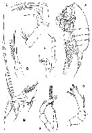 Issued from : G.A. Boxshall & S.H. Halsey in An Introduction to Copepod Diversity. The Ray Society, 2004, No 166, Part. I. [p.85, Fig. 12]. Candaciidae. A, Candacia norvegica female; B, Candacia longimana (as Candace longimana) female Mx1; C, Mx2; D, Candacia pachydactila (as Candace pachydactyla) female P5; E, Candacia simplex (as Candace simplex) male P5; F, Candacia catula (as Candace catula maleP5; G, Candacia armata (as Candace pectinata) female P4 endopod.
Sars, 1902 (A); Giesbrecht, 1893 a (B-G). |
 Issued from ; J.M. Bradford-Grieve in NIWA Biodiv. Mem. ( 1999, p.160]. Seta and spine formula of the swimming legs P1 to P4. - P5 female uniramous, not natatory, usually symmetrical; coxa and coupler (intercoxal sclerite) fused; basis and 1-segmented exopod separate, with 1 distolateral spinule; terminal segment longest, either may end in one or more spin-like processes; a finger-like process, or a single long seta; setae may or may not be present on the inner margins. - P5 male not natatory, 4-segmented on left and 3-segmented on right; may be chelate on right or ending in a long feather-like seta; coxa not counted in segmental complement, are small and fused with the coupler. | | | | | | Syn.: | Candace : Dana,1849; 1852; Claus, 1863 (p.189); Brady, 1878 (p.48); Giesbrecht, 1892 (p.67, 423); Wheeler, 1901 (p.177); Ifionyx Kröyer, 1845; 1849 (in Damkaer & Damkaer, 1979, p.35);
Paracandacia : Grice, 1963 (p.172); Owre & Foyo, 1967 (p.95); Bradford,1972 (p.30); Björnberg & al., 1981 (p.655); Razouls, 1982 (p.548); Mauchline, 1988 (p.713); Razouls, 1993 (p.307); Chihara & Murano, 1997 (p.755); Mulyadi, 1997 (p.102); Mauchline, 1998 (p.95); Vaupel Klein & Gassmann, 1998 (p.441, Rem.); Bradford-Grieve & al., 1999 (p.955, spp. Key in South Atlantic); Bradford-Grieve, 1999 b (p.176, Def.); Mulyadi, 2004 (p.76, spp. Key in Indonesian waters); Vives & Shmeleva, 2007 (p.460, spp. Key).
Candacia spp.: Webber & Roff, 1995 a (p.481, Tableb 1, 2, 3, biomass & production) | | Ref.: | Giesbrecht & Schmeil, 1898 (p.126, spp. Key); Sars, 1902 (1903) (p.133); Esterly, 1905 (p.192, spp. Key); van Breemen, 1908 a (p.145, spp. Key); A. Scott, 1909 (p.150); Sewell, 1932 (p.334); Wilson, 1932 a (p.138, spp. Key); Rose, 1933 a (p.248, spp. Key); Mori, 1937 (1964) (p.78, spp. Key); Pesta, 1941 (p.158); Farran, 1948 b (n°13, p. 3); Davis, 1949 (p.61); Brodsky, 1950 (1967) (p.403); Grice & Vervoort, 1963 (p.150); Grice, 1963 (p.173, Rev., spp. Key); Crisafi, 1963 (p.81); Grice, 1963 a (p.173); Tanaka, 1964 c (p.243); Chen & Zhang, 1965 (p.88, spp. Key); Owre & Foyo, 1967 (p.91, spp. Key); Lawson, 1973 (p.302, Rem.); 1977 (p.76, Rem.); Björnberg & al., 1981 (p.655, clé. spp.); Razouls, 1982 (p.535); Gardner & Szabo, 1982 (p.403); Zheng Zhong & al., 1984 (1989) (p.250, clé spp.); Mauchline, 1988 (p.713); Huys & Boxshall, 1991 (p.461); Razouls, 1993 (p.307); Mulyadi, 1997 a (p.68, spp. Key); Chihara & Murano, 1997 (p.752, spp. Key); Mauchline, 1998 (p.95); Vaupel Klein & Gassmann, 1998 (p.441, Rem.: phylogénie); Bradford-Grieve & al., 1999 (p.955, 956: clé spp.); Bradford-Grieve, 1999 b (p.161, Déf.); Mulyadi, 2004 (p.79, Def., spp. Key in Indonesian waters); Vives & Shmeleva, 2007 (p.438, spp. Key). | | Rem.: | type: C. ornata in Fowler (1912), replaced by the species type
Candacia pachydactyla (Dana,1849) in Grice & Vervoort, 1963 (p.150). 32 spp. + 3 unidentified (+ 2 doubtful : guinensis, inermis).
Key to species of Paracandacia and Candacia after Bradford-Grieve & al. (1999, p.955-957) from South Atlantic :
Female :
0 P5 terminal segments end in processes at least one of which is finger-like process and may be finely serrated on one or both margins
.. Paracandacia (= Candacia).
0 P5 terminal segments end in one or more spine-like processes
. Candacia.
01 - In dorsal view, genital segment with spine-like protrusions arising from each side and directed posteriorly (Fig.7.375, p.1066)
Paracandacia bispinosa (= Candacia bispinosa)
01 In dorsal view genital segment without spine-like protrusions
02.
02 - P5 terminal finger-like process finely serrate proximally ; distal seta on inner margin approximately twice as long as proximal seta ; points of posterior corners of prosome directed backwards (Fig.7.376, p.1066)
.. Paracandacia simplex (= Candacia simplex).
02 - P5 terminal finger-like process finelly serrate distally ; setae on inner margin subequal, distal seta slightly longer than proximal seta ; points at posterior corners of prosome directed ventrally, scarcely visible in dorsal view (Fig.7.377, p.1066)
Paracandacia truncata (= Candacia truncata)
0 : Candacia
1 Posterior corners of prosome broadly rounded (Fig.7.366, p.1065)
. Candacia elongata.
1 Posterior corners of prosome pointed or otherwise produced
2.
Urosomal segment 2 with ventral protrusion, lamella, or spuine-like process arising from ventral surface
3.
2 Urosomal segment 2 without lateral or ventral protrusion and without spine-like process arising from ventral surface
. 6.
3 In dorsal view, lateral margins of genital segment somewhat triangula rand pointed ; lamella on ventral surface of Urosomal segment 2 (Fig.7.362, p.1064)
.. Candacia bipinnata.
3 In dorsal view, lateral margins of genital segment not pointed
4.
4 In dorsal view, protrusion on ventral surface of urosomal segment 2 visible on right side of somite ; in dorsal view, lateral margins of genital segment with knob-like protrusions ; apex of P5 ends in long saber-like point (Fig.7.361, p.1064)
Candacia armata4 In dorsal view, protrusion on ventral surface of urosomal segment 2 not visible ; apex of P5 ends in 2 or more points
.. 5.
5 In lateral view, ventral protrusion of urosomal segment 2 directed obliquely anteriad ; terminal segment of P5 with 2 small, outer, spine-like points and 2 small, subequal, spine-like points at tip (Fig.7.372, p.1065)
.. Candacia paenelongimana.
5 In lateral view, ventral protrusion of urosomal segment 2 directed obliquely posteriad ; terminal segment of P5 with 2 small, outer, spine-like points and 3 spine-like points distally, middle one of which is longest (Fig.7.364, p.1064)
Candacia cheirura.
6 No inner edge setae present on terminal segment of P5
7.
6 2 or 3
inner edge setae present on terminal segment of P5
11.
7 Spine-like process present on ventral side of genital segment (Fig.7.365, p.1064)
.. Candacia curta.
7 No spine-like process present on ventral side of genital segment
. 8.
8 3 spine-like points present on terminal segment of P5 (Fig.7.373, p.1066)
.. Candacia tenuimana.
8 4 or 5 spines or spine-like points present on terminal segment of P5
. .
9 Apex of terminal segment of P5 with 3 subequal spine-like points (Fig.7.368, p.1065)
Candacia longimana.
9 Apex of terminal segment of P5 with 1 spine-like points or 2 unequal spine-like points
. 10.
10 Terminal segment of P5 with 2 outer spines and 2 distal spines (Fig.7.374, p.1066)
.. Candacia varicans.
10 Terminal segment of P5 with 3 small outer spines, and 2 distal spines (outer spine very small, inner spine large) (Fig.7.369, p.1065)
.. Candacia magna.
11 No spines or spine-like processes present on genital segment ; in dorsal view, genital segment with distinctly convex protrusion on each side ; in lateral view, genital segment with ventral, rounded protrusion directed posteriad (Fig.7.363, p.1064)
Candacia catula.
11 Spines or spine-like processes present on dorsal, lateral or ventral margins of genital segment
12.
12 In dorsal view, 1 robust spine-like process extending obliquely posteriad from left side, and 1 robust spine extending posteriad from right side of genital segment ; both surpass posterior margin of genital segment (Fig.7.371, p.1065)
.. Candacia pachydactyla.
12 In dorsal view, spines from left and right sides of genital segment smaller and not reaching posterior margin of genital segment
13.
13 In lateral view, small protuberance arising from ventral side of genital segment near posterior margin ; distal 2 inner setae on terminal segment of P5, coaese and ubequal in length (Fig.7.367, p.1065)
.. Candacia ethiopica.
13 In lateral view, no protuberance on ventral sutface og genital segment ; distal 2 inner setae on terminal segment of P5, thin and approximatelt equal in length (Fig.7370, p.1065)
.. Candacia norvegica.
Male : :
0 - P5 is not chelate on right but ends in long plumose seta ; right A1 has no teeth in geniculate region
.. Paracandacia .
0 P5 is chelate on right ; right A1 has teeth on one or more segments in geniculate region
. Candacia.
- 01 Right A1 segments 16 and 19-20 without rounded or elongate protrusions (Fig. 7.376, p.1066)
.. Paracandacia simplex (= Candacia simplex).
01 - One or both of right A1 segments 16 and 19-20 with rounded or elongate protrusions
. 02.
02 A1 segment 16 with rounded protrusion distally, segment 19-20 produced distally (Fig. 7.375, p.1066)
. Paracandacia bispinosa (= Candacia bispinosa-.
02 - A1 segment 16 with elongate protrusion distally, segment 19-20 not produced (Fig. 7.377, p.1066)
. Paracandacia truncata (= Candacia truncata).
01 Left posterior corner of prosome rounded (Fig. 7.366, p.1065)
Candacia elongata.
01 Left posterior corner of the prosome pointed
02
02 In dorsal view, genital segment without process or protrusion
03.
02 In dorsal view, genital segment with process or protrusion
. 04.
03 Proximal spine on Mx2 inner lobe 5 considerably thicker than distal spine (Fig. 7.363, p.1064)
Candacia catula.
03 Proximal spine on Mx2 inner lobe 5 not notably thicker than distal spine (Fig. 7.374, p.1066)
Candacia varicans.
04 Right A1 with segments 2 and 3 fused
.. 05.
04 Right A1 with segments 2 and 3 separate
11.
05 Right A1 with segments 17 and 18 fused
6.
05 Right A1 with segments 17 and 18 separate
.. 7.
06 In dorsal view, process on right side of genital segment small, consisting of rounded knob in front of which is pointed projection (Fig. 7.367, p.1065)
.. Candacia ethiopica.
06 In dorsal view, process on right side of genital segment large, consisting of single broad and rounded projection (Fig. 7.371, p.1065)
.. Candacia pachydactyla.
07 Right A1 with segments 19 and 20 fused or partially fused
.. 08.
07 Right A1 with segments 19 and 20 separate
010.
08 In lateral view, distal end of right posterior corner of prosome truncate, extending beyond posterior border of genital segment (Fig. 7.362, p.1064)
. Candacia bipinnata.
08 In lateral view, distal end of right posterior corner of prosome not truncate, may or may not extend beyond posterior border of genital segment
09.
09 In lateral view, right posterior prosome corner not reaching posterior border genital segment and turned upwards (Fig. 7.365, p.1064)
Candacia curta.
09 In lateral view, right posterior prosome corner reaching posterior border of genital segment and turned slightly downwards Fig. 7.361, p.1064)
.. Candacia armata.
010 - Tip of thumb of chela on right P5 reaching to about midlength of finger (Fig. 7.361, p.1064)
. Candacia armata.
010 Tips of thumb and finger of right P5 subequal (Fig. 7.369, p.1065)
.. Candacia magna.
011 In dorsal view, distal end of process on right side of genital segment ending in point
. 012.
011 In dorsal view, distal end of process on right side of genital segment rounded or lobate
014.
012 Distal segment of left P5 longer than penultimate segment (Fig.7.364, p.1064)
.. Candacia cheirura.
012 Distal segment of left P5 shorter than penultimate segment
. 013.
013 In dorsal view, process on genital segment directed outwards with distal end curved posteriorly ; right posterior corner of prosome notched at tip (Fig.7.373, p.1066)
Candacia tenuimana.
013 In dorsal view, process on genital segment not curved and directed somewhat obliquely posteriad ; right posterior prosome corner not notched at tip (Fig. 7.372, p.1065)
. Candacia paenelongimana.
014 In dorsal view, process on genital segment divided into 2 lobes (Fig. 7.370, p.1065)
.. Candacia norvegica.
014 In dorsal view, process on genital segment knob-like Fig. 7.368, p.1065)
. Candacia longimana
After Ohtsuka & Nishida (2017, p.581), Candacia is specialized for predation on gelatinous zooplankters such as appendicularians (Ohtsuka & Onbé, 1989; Ohtsuka & Kubo, 1991)..
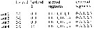
| | Remarks on dimensions and sex ratio: | | The mean female size is 2.530 mm (n = 63; SD = 0.9220), and the mean male size is 2.416 mm (n = 62; SD = 0.8998). The size ratio (male: female) is 0.955. The sex ratio (Female: male) is 1. | | | | Table I: Candacia : Total length, localization and depth
Species |
Lg (mm) F; M |
Localization |
Depth |
C. grandis |
4 à 5 |
Japan |
|
C. magna |
3 à 4,5 |
Atlant- Tropic. Indian |
|
C. columbiae |
- |
N. Pacif.N |
|
C. parafalcifera |
- |
N. Pacif N, Pacif E (bathypel) |
|
C. falcifera |
3 à 4 |
Antarct., Groenland |
|
C. maxima |
- |
Antarct, sub-Antarct + C.S.T |
|
C. elongata |
2 à 3,9 |
Cosmop. Tropic |
M - B |
C. longimana |
- |
Cosmop. Tropic |
E- M |
C. norvegica |
- |
Cosmop. cold temperate |
E- M |
C. pachydactila |
> 2 à > 3,5 |
Cosmop. Tropic |
E - B |
C. armata |
2 à 3 |
warm & cold temperate |
E - M |
C. bibinnata |
- |
Cosmop. Tropic, cold temperate |
E - M |
C. cheirura |
- |
Sub-Antarct + CST |
E - M |
C. curta |
- |
Cosmop. Tropic |
E - B |
C. ethiopica |
- |
Cosmop. Tropic |
E - M |
C. giesbrechti |
- |
warm temperate |
|
C. ishimarui |
- |
Tropic. |
|
C. paenelongimana |
- |
Tropic. |
M |
C. pofi |
- |
Tropic. |
|
C. samassae |
- |
Tropic. |
|
C. tenuimana |
- |
Tropic. |
M |
C. varicans |
- |
Cosmopol. Tropic |
E - M |
C. simplax |
- |
Cosmopol. Tropic |
E - M |
C. truncata |
- |
Cosmopol. Tropic |
|
C. bradyi |
1 à 2 |
Tropic. |
|
C. catula |
- |
Tropic. |
E |
C. discaudata |
- |
Tropic. |
|
C. guggenhemi |
- |
Tropic. |
|
C. ketchumi |
- |
Tropic, sub-Tropic. |
E - M |
The largest form, C. grandis , was described from Japan, the smallest, C. catula, from the Indo-Pacific.
The largest forms correspond to cold waters (Antarctic or N. Pacific), except C. magna.
Four species show highest deviations between minimum and maximum values. These correspond to tropical or subtropical cosmopolite forms and generally meso- or bathypelagic. Fourteen species (being 44%) have sizes between 2 and 3 mm, for females as well as for males, which are in general slightly smaller than the females. Eight species show smaller dimensions, between 1 and 2 mm, and are observed in tropical waters.
| | | | | Ref.: | Grice, 1963 (p.172); Owre & Foyo, 1967 (p.95); Bradford,1972 (p.30); Björnberg & al., 1981 (p.655); Razouls, 1982 (p.548); Mauchline, 1988 (p.713); Razouls, 1993 (p.307); Chihara & Murano, 1997 (p.755); Mulyadi, 1997 (p.102); Vaupel Klein & Gassmann, 1998 (p.441, Rem.); Bradford-Grieve & al., 1999 (p.955, clé spp.); Bradford-Grieve, 1999 b (p.176, Déf.) | | Rem.: | This genus is considered as non-valid by Boxshall & Halsey (2004, p.84).
Cf. Candacia | | | | |
|
|
 Any use of this site for a publication will be mentioned with the following reference : Any use of this site for a publication will be mentioned with the following reference :
Razouls C., Desreumaux N., Kouwenberg J. and de Bovée F., 2005-2025. - Biodiversity of Marine Planktonic Copepods (morphology, geographical distribution and biological data). Sorbonne University, CNRS. Available at http://copepodes.obs-banyuls.fr/en [Accessed August 27, 2025] © copyright 2005-2025 Sorbonne University, CNRS
|
|
|
|


;)
