|
|
 |
|
Calanoida ( Order ) |
|
|
|
Diaptomoidea ( Superfamily ) |
|
|
|
Candaciidae ( Family ) |
|
|
|
Candacia ( Genus ) |
|
|
| |
Candacia truncata (Dana, 1849) (F,M) | |
| | | | | | | Syn.: | Candace truncata Dana, 1849; Giesbrecht, 1892 (p.425, 440, 771, figs.F,M); no Brady, 1883 (p.69, figs.F,M); ? T. Scott, 1894 b (p.63);
Candacia turgida C.B. Wilson, 1950 (p.183, figs.F); Ganapati & Shanthakumari, 1962 (p.9, 15);
Paracandacia truncata : Grice, 1963 (p.173, fig.F,M, Rem.); Vidal, 1968 (p.43, figs.F,M); Corral Estrada, 1970 (p.203: Rem.); Lawson, 1977 (p.71, tab.2,3,4, fig.4, 5); Carter, 1977 (1978) (p.36); Greenwood, 1978 (p.11, figs.F,M); Chihara & Murano, 1997 (p.755, Pl.79,80: F,M); Mulyadi, 1997 a (p.106, figs.FM, Rem.); Vaupel Klein & Gassmann, 1998 (p.443, fig.M); Bradford-Grieve & al., 1999 (p.885, 955, figs.F,M); Bradford-Grieve, 1999 b (p.177, figs.F,M, figs.183,193); Conway & al., 2003 (p.114, figs.F,M, Rem.); Mulyadi, 2004 (p.103, figs.F,M, Rem.); Phukham, 2008 (p.64, figs.F,M);
Ref. compl.: Grice & Hulsemann, 1967 (p.19); Fleminger, 1967 a (tabl.1); Morris, 1970 (p.2301); Dessier, 1983 (p.89, Tableau 1, Rem., %); Guangshan & Honglin, 1984 (p.118, tab.); Almeida Prado Por, 1985 (p.250); Madhupratap & Haridas, 1986 (p.105, tab.1); Dessier, 1988 (tabl.1); Madhupratap & Haridas, 1990 (p.2305, fig.4: vertical distribution night/day; fig.7: cluster); Othman & al., 1990 (p.561, 564, Table 1); Yoo, 1991 (tab.1); Ohtsuka & Kubo, 1991 (p.538); Shih & Young, 1995 (p.69); Padmavati & Goswami, 1996 a (p.85, fig.3); Padmavati & al., 1998 (p.349); Madhupratap & al., 2001 (p. 1345, vertical distribution vs. O2, figs.4, 5: clusters); Lo & al., 2001 (1139, tab.I); Hsiao & al., 2004 (p.325, tab.1); Lan & al., 2004 (p.332, tab.1); Lo & al.*, 2004 (p.218, fig.6); Yin & al., 2004 (p.3); Lo & al., 2004 (p.89, tab.1); Osore & al., 2004 (p.195); Kazmi, 2004 (p.228); Lavaniegos & Jiménez-Pérez, 2006 (p.150, tab.2, 3, Rem.); Zuo & al., 2006 (p.162: tab.1); Hwang & al., 2006 (p.943, tabl. I); Hwang & al., 2007 (p.23); Dur & al., 2007 (p.197, Table IV); Jitlang & al., 2008 (p.65, Table 1); Fernandes, 2008 (p.465, Tabl.2); Ayon & al., 2008 (p.238, Table 4: Peruvian samples); Lan Y.C. & al., 2008 (p.61, Table 1, % vs stations); C.-Y. Lee & al., 2009 (p.151, Tab.2); Tseng L.-C. & al., 2008 (p.153, Table 2, occurrence vs geographic distribution); Tseng & al., 2009 (p.327, fig.5, feeding); Lan Y.-C. & al., 2009 (p.1, Table 2, % vs hydrogaphic conditions); Fazeli & al., 2010 (p.153, Table 1); W.-B. Chang & al., 2010 (p.735, Table 2, abundance); Xu & Gao, 2011 (p.514, figs.3, 4, Table 2: optimal salinity); Guo & al., 2011 (p.567, table 2, indicator); Hsiao & al., 2011 (p.317, Table 2, fig.6, indicator of seasonal change); Tseng L.-C. & al., 2011 (p.47, Table 2, occurrences vs mesh sizes); Tseng & al., 2012 (p.621, Table 3: abundance); Tseng & al., 2013 (p.507, seasonal abundance); Tseng & al., 2013 a (p.1, Table 3, 4, abundance); Hirai & al., 2013 (p.1, Table I, molecular marker); Hwang & al., 2014 (p.43, Appendix A: seasonal abundance)
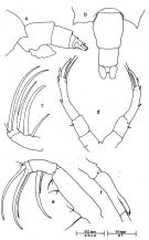
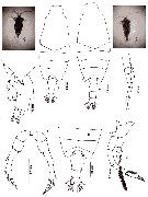
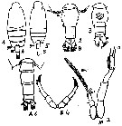
Canadacia truncata : Go & al., 1997 (tab.1: lapsus calami) | | | | Ref.: | | | Thompson, 1888 d (p.140); Giesbrecht & Schmeil, 1898 (p.130, Rem. F); Thompson & Scott, 1903 (p.235, 250); Cleve, 1904 a (p.187); A. Scott, 1909 (p.155, Rem.); Farran, 1936 a (p.115); Dakin & Colefax, 1933 (p.207); Tanaka, 1935 a (p.214, figs.F,M, juv.); Mori, 1937 (1964) (p.82, figs.F,M); Dakin & Colefax, 1940 (p.103, figs.F,M); Pesta, 1941 (p.169, figs.F,M); Wilson, 1942 a (p.175, fig.F); Krishnaswamy, 1953 (p.129); Chiba, 1956 (p.63, figs.F,M); Chiba & al., 1957 (p.310); 1957 a (p.11); Grice & Jones, 1960 (p.288, figs.F, Rem.); Brodsky, 1962 c (p.137, figs.F,M); Grice, 1962 (p.239, figs.F,M); Fagetti, 1962 (p.34); Tanaka, 1964 c (p.245); De Decker, 1964 (p.15, 19, 27); De Decker & Mombeck, 1964 (p.11); Chen & Zhang, 1965 (p.91, figs.F,M); Saraswathy, 1966 (1967) (p.81); Delalo, 1968 (p.138); Itoh, 1970 (tab.1); Kos, 1972 (Vol.I, figs F, M, Rem.); Tranter, 1977 (p.596, 601); Frontier, 1977 a (p.16); Chen Q-c, 1980 (p.794); De Decker, 1984 (p.317, 337: carte); M. Lefèvre, 1986 (p.33); Renon, 1987 (tab.2); Hernandez-Trujillo, 1989 a (tab.1); Cervantes-Duarte & Hernandez-Trujillo, 1989 (tab.3); Hernandez-Trujillo, 1991 (1993) (tab.I); Kim & al., 1993 (p.268); Park & Choi, 1997 (Appendix); Hwang & al., 1998 (tab.II); Wong & al, 1998 (tab.2); Suarez-Morales & Gasca, 1998 a (p109); Hernandez-Trujillo, 1999 (p.284, tab.1); Lavaniegos & Gonzalez-Navarro, 1999 (p.239, Appx.1); Dolganova & al., 1999 (p.13, tab.1); Fernandez-Alamo & al., 2000 (p.1139, Appendix); Suarez-Morales & al., 2000 (p.751, tab.1); Hwang & al., 2003 (p.193, tab.2); Hernandez-Trujillo & Esqueda-Escarcega, 2002 (in Appendix); Hernandez-Trujillo & Suarez-Morales, 2002 (p.748, tab.1); Gallienne & al., 2004 (p.5, tab.3); Boxshall & Halsey, 2004 (Rem. p.84) | 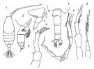 issued from : O. Tanaka in Suisan Gakkai Ho, Tokyo, 1935, 6 (4). [Pl.VI, p.227]. Female: 1, habitus (dorsal view); 2, last thoracic segment and urosome (lateral view); 3, idem (from other specimen); 4, P5. Male: 7, last thoracic segment and urosome (dorsal view); 8, right A1 (hinge segments); 9, P5 (left and right)
|
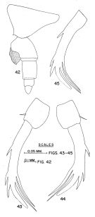 issued from: G.D. Grice & E.C. Jones in Pacific Science, 1960, XIV (3). [p.287, Figs.42-45]. Female (from central Pacific): 42, 4th and 5th thoracic segments and urosome (lateral left side), showing cement mass attached to genital segment; 43, 44, P5 (unlike from same specimen); 45, P5 (with serrated external spinous processes from another specimen). Nota: The mass of cement associated with a spermatophore is present on some females.
|
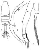 issued from: Q.-c Chen & S.-z. Zhang in Studia Marina Sinica, 1965, 7. [Pl.37, 1-3]. Male (from E China Sea): 1, habitus (dorsal); 2, right A1 (16th-19th segments); 3, P5 (posterior).
|
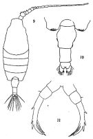 issued from: Q.-c Chen & S.-z. Zhang in Studia Marina Sinica, 1965, 7. [Pl.36, 9-11]. Female: 9, habitus (dorsal); 10, postero-lateral angles of last thoracic segment and urosome (dorsal); 11, P5 (posterior).
|
 issued from : J.G. Greenwood in Proc. R. Soc. Qd, 1978, 89. [p.12, Fig.5]. As Paracandacia truncata. Female (from Moreton Bay, E Australia): a, posterior part of metasome and urosome (lateral left side); b, idem (dorsal); c, Mx2; d, P5. Male: e, Mx2; f, right A1 (segment 16). Nota: Elongate lobe developed on segment 16 of right A1.
|
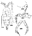 issued from : C.B. Wilson in Bull. U.S. natn. Mus., 1950, 100, 14 (4). [Pl. 22, Figs.305-308]. As Candacia turgida. Female (from 14°45'N, 120°12'30''E): 305,habitus (dorsal); 306,posterior prosome and urosome (right lateral); 307,proximal segments of A1; 308, P5.
|
 issued from : T. Mori in The Pelagic copepoda from the neighbouring waters of Japan, 1937 (1964). [Pl. 55, Figs.1-6.]. Female: 1, habitus (dorsal); 2, Mx2; 3, last thoracic segment (right side); 6, P5.. Male: 4, habitus (dorsal); 5, P5 (posterior);
|
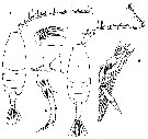
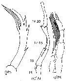
 issued from : G.D. Grice in Fish. Bull. Fish and Wildl. Ser., 1962, 61. [p.237, Pl.33, Figs.6-15]. Female (from equatorial Pacific): 6-7, habitus (dorsal and lateral, respectively); 8, Md (biting edge); 9, Mx2; 10, P5. Nota: Genutal segment symmetrical and finely pubescent on the lateral margins. The terminal ginger of the distal segment of P5 is finely serrate along its outer distal margin; the proximal seta on the internal margin is slightly shorter than the distal one. Male: 11-12, habitus (dorsal and lateral, respectively); 13, posterior part of thorax and urosome (dorsal); 14, segments 15 to 20 of right A1; 15, P5. Thorax and urosome symmetrical. Segment 16 of right A1 produced into an elongate process The distal spine on the terminal segment of right P5 is considerably larger than the preceding two spines; the terminal seta on some specimens is articulated to this segment while in others it is not.
|
 issued from : N. Phukham in Species diversity of calanoid copepods in Thai waters, Andaman Sea (Master of Science, Univ. Bangkok). 2008. [p.147, Fig.21]. As Paracandacia truncata. Female (from W Malay Peninsula): a, habitus (dorsal); b, urosome; c, urosome (lateral, left side); d, P5. Male: e, habitus (dorsal); f, segments 15 and 16 of A1; g, P5. Body length after the drawings: F = 1.889 mm; M = 1.979 mm.
|
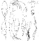
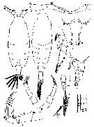 issued from : Mulyadi in Published by Res. Center Biol., Indonesia Inst. Sci. Bogor, 2004. [p.104, Fig.58]. As Paracandacia truncata. Female (from 06°10'S, 106°00'E): A, habitus (dorsal); B-C, posterior part of last thoracic segment and urosome (dorsal and lateral, respectively); D, Mx2; E, P3; F, P5. Male: G, habitus (dorsal); H, geniculate region of right A1; I, P5.
|
 Issued from : J.M. Bradford-Grieve, E.L. Markhaseva, C.E.F. Rocha & B. Abiahy in South Atlantic Zooplankton, edit. D. Boltovskoy. 1999, Vol. 2, Copepoda; [p.1066, Fig. 7.377: Candacia truncara (as Paracandacia trubcata)]. r = right. Female characters (from key, p.955): - P5 terminal segments end in processes at least one of which is finger-like process and may be finely serrated on one or both margins. - In dorsal view, genital segment without spine-like protrusions. - P5 terminal finger-like process finely serrate distally; setae on inner margin subequal, distal seta slightly longer than proximal seta; points at posterior corners of prosome directed ventrally, scarcely visible in dorsal view Male (from key, p.925): - P5 not chelate on right but ends in long plumose seta; right A1 has no teeth in geniculate region. - One or both of right A1 segments 16 and 19-20 with rounded or elongate protrusions. - A1 segment 16 with elongate protrusion distally, segment 19-20 not produced.
|
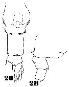 Issued from : W. Giesbrecht in Fauna Flora Golf. Neapel, 1892, 19. [Taf. 39, figs. 26, 28]. As Candace truncata. Female: 26, urosome (ventral); 28, thoracic segment 5 and abdominal segments 1 and 2.
|
 Issued from : M.C. Kos in Field guide for plankton. Zool. Institute USSR Acad., Vol. I, 1972. As Paracandacia truncata. After Brodsky, 1962. Female: 1, habitus (dorsal); 2, corner of the last thoracic segment and abdomen (dorsal); 3, abdomen (ventral); 4, P5. Male: 5, habitus (dorsal); 6, corner of the last thoracic segment and abdomen (dorsal); 7, P5.
| | | | | Compl. Ref.: | | | Carl, 1907 (p.17); Sewell, 1912 (p.353, 368); 1914 a (p.230); 1932 (p.338); 1948 (p.392, 396, 408, 412, 415, 423, 433, 443, 453, 457, 460); Yamazi, 1958 (p.151, Rem.); Ahlstrom & Thrailkill, 1963 (p.57, Table 5, abundance); Timonin, 1971 (p.281, trophic group); Stephen, 1984 (p.161, 169, Distribution vs thermocline & geographic); Brinton & al., 1986 (p.228, Table 1); Chen Y.-Q., 1986 (p.205, Table 1: abundance %, Table 2: vertical distribution); Noda & al., 1998 (p.55, Table 3, occurrence); McKinnon & al., 2008 (p.843: Tab.1); Hernandez-Trujillo & al., 2010 (p.913, Table 2); Hsiao S.H. & al., 2011 (p.475, Appendix I); Maiphae & Sa-ardrit, 2011 (p.641, Table 2); Tutasi & al., 2011 (p.791, Table 2, abundance distribution vs La Niña event); Uysal & Shmeleva, 2012 (p.909, Table I); in CalCOFI regional list (MDO, Nov. 2013; M. Ohman, comm. pers.); Zakaria & al., 2016 (p.1, Table 1, Rem.); Jerez-Guerrero & al., 2017 (p.1046, Table 1: temporal occurrence); Abo-Taleb & Gharib, 2018 (p.139, Table 5, occurrence %); Palomares-Garcia & al., 2018 (p.178, Table 1: occurrence) | | | | NZ: | 11 + 2 doubtful | | |
|
Distribution map of Candacia truncata by geographical zones
|
| | | | | | | | | | | | 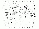 issued from: T. J. Lawson in Marine Biology, 1977, 43. [Fig.5, p.77]. As Paracandacia truncata issued from: T. J. Lawson in Marine Biology, 1977, 43. [Fig.5, p.77]. As Paracandacia truncata
Distribution map for the Indian Ocean. |
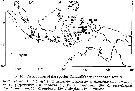 issued from : Mulyadi in Treubia, 1997, 31 (2). [p.109, Fig.16]. issued from : Mulyadi in Treubia, 1997, 31 (2). [p.109, Fig.16].
Distribution of Candaciidae in Indonesian waters. 14: C. truncata. |
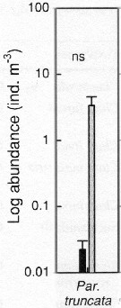 Issued from : S.-H. Hsiao, S. Kâ, T.-H. Fang & J.-S. Hwang inHydrobiologia, 2011, 666. [p.326, Fig.6]. Issued from : S.-H. Hsiao, S. Kâ, T.-H. Fang & J.-S. Hwang inHydrobiologia, 2011, 666. [p.326, Fig.6].
Variations in the most abundant copepod species (mean ± SE) along the transect in the boundary waters between the northern part Taiwan Strait and the East China Sea i March (black bar) and October (grey bar) 2005 (Mann-Whitney U test, sig. ns: P >0.05.
See drawings of Hydrological conditions and superficial marine currents in Calanus sinicus. |
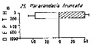 Issued from : M. Madhupratap & P. Haridas in J. Plankton Res., 12 (2). [p.311, Fig.4]. Issued from : M. Madhupratap & P. Haridas in J. Plankton Res., 12 (2). [p.311, Fig.4].
Vertical distribution of calanoid copepod (mean +1 SE), abundance No/100 m3. 25- Paracandacia (= Candacia) truncata.
Night: shaded, day: unshaded.
Samples collected from 6 stations located off Cochin (India), SE Arabian Sea, November 1983, with a Multiple Closing Plankton Net (mesh aperture 300 µm), in vertical hauls at 4 depth intervalls (0-200, 200-400, 400-600, 600-1000 m). |
| | | | Loc: | | | South Africa (E & W), ? G. of Guinea, E Mditerranean Sea (Malta, W Egyptian coast, Lebanon Basin), G. of Elat, Hurghada (shallow sheltered lagoon), Red Sea, Gulf of Oman, G. of Aden, Arabian Sea, Maldive Is., Sri Lanka, Madagascar (Tuléar, Nosy Bé), Rodrigues Is. - Seychelles, Kenya, Natal, India (Goa, Lawson's Bay, Madras), Bay of Bengal, W Malay Peninsula (Andaman Sea), Australia (W, North West Cape), Indonesia-Malaysia, Jakarta-Seribu Islands, Flores Sea, Ambon Bay, S Celebes Sea, Philippines, Hainan (Sanya Bay), Hong Kong, China Seas (Yellow Sea, East China Sea, South China Sea), Taiwan Strait, Taiwan (S, E, SW, NW, N, NE), Okinawa, S Korea, Japan Sea, Japan, Kuchinoeranu Is., Izu region, Tanabe Bay, off SE Japan, Bikini, W Pacif. (equatorial), Australia (G. of Carpentaria, Great Barrier, Moreton Bay, New South Wales), New Caledonia, California, W Baja California, Bahia Magdalena, G. of California, Pacif. (equatorial), Moorea Is., off N Hawaï, W Mexico, La Paz, G. de Tehuantepec, Galapagos-Ecuador, Bahia Cupica (Colombia), Peru, Chile | | | | N: | 135 (Atlant. S: 1; Medit.: 2; Red Sea: 6; Indian: 37; Indonesia-Malaysia: 7; Pacif.: 82) | | | | Lg.: | | | (34) F: 2,04-1,85; (46) F: 2-1,85; M: 2-1,9; (101) F: 2,04-1,84; M: 2,11-1,87; (104) F: 2,3; M: 2,1; (120) F: 2,23-2,02; M: 2,15; (137) F: 2,3-2,1; (150) F: 2,17-1,9; M: 2,12-1,97; (187) F: 2,18; M: 2,18-2,06; (290) F: 2,1-1,9; M: 2,25-1,95; (530) ?: 1,4; (778) F: 1,82-1,75; M: 2-1,95; (786) F: 2,1-1,78; M: 2,05; (795) F: 1,5; M: 1,3; (991) F: 1,84-2,1; M: 1,87-2,11; (1122) F: 1,75; M: 1,95; (1230) F & M: 1,9-2,2; {F: 1,50-2,30; M: 1,30-2,25}
The mean female size is 1.978 mm (n = 24; SD = 0.1976), and the mean male size 1.994 mm (n = 19; SD = 0.1991). The size ratio (male : female) is 1.012 (n = 12; SD = 0.0709). | | | | Rem.: | The indications from the Atlantic (Gulf of Guinea, Canaries, Madeira, Faroe Islands) and in the Mediterranean are considered as very doubtful and need confirmation.
For Greenwood (1978, p.11) an oceanic form rarely taken inshore waters.
After Itoh (1970 a, fig.2, co-ordinates) the Itoh's index value of the mandibular gnathobase = 2640 (as Paracandacia truncata).
Timonin (1971, p.282) considers the trophic interrelations in the equatorial and tropical Indian Ocean, and divides the plankters into 6 trophic groups from the litterature and the results of studies of mouth-parts structure and intestine content. This species is a piercing and sucking carnivorous.
See in DVP Conway & al., 2003 (version 1) | | | Last update : 19/06/2023 | |
|
|
 Any use of this site for a publication will be mentioned with the following reference : Any use of this site for a publication will be mentioned with the following reference :
Razouls C., Desreumaux N., Kouwenberg J. and de Bovée F., 2005-2025. - Biodiversity of Marine Planktonic Copepods (morphology, geographical distribution and biological data). Sorbonne University, CNRS. Available at http://copepodes.obs-banyuls.fr/en [Accessed January 05, 2026] © copyright 2005-2025 Sorbonne University, CNRS
|
|
|
|


;)
;)
;)
;)
;)
;)
;)
;)
;)
;)
;)
;)
;)

;)
;)
;)
;)
