|
|
 |
|
Calanoida ( Order ) |
|
|
|
Clausocalanoidea ( Superfamily ) |
|
|
|
Scolecitrichidae ( Family ) |
|
|
|
Scolecitrichopsis ( Genus ) |
|
|
| |
Scolecitrichopsis tenuipes (T. Scott, 1894) (F,M) | |
| | | | | | | Syn.: | Scolecithrix tenuipes T. Scott, 1894 b (p.48, Descr.M, figs.M); Giesbrecht & Schmeil, 1898 (p.47, Rem.M); Thompson & Scott, 1903 (p.234, 245); Bradford & al., 1983 (p.104);
Scolecithricella tenuipes : A. Scott, 1909 (p.92, Rem.M); Sewell, 1948 (p.516, 517, 523, 546); Bainbridge, 1972 (p.61, Appendix Table I: vertical distribution vs day/night); Dessier, 1979 (p.205);
Scolecithricella (Amallothrix) marquesae Vervoort, 1965 (p.74, Descr.F, figs.F, Rem.);
Amallothrix sp. Marques, 1958 (p.212);
Amallothrix marquesae : Vives, 1982 (p.292);
Scolecithricella (Amallothrix) marquesae : Vervoort, 1965 (p.79, figs.F);
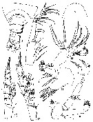
Scolecithricella marquesae : Binet & al., 1972 (p.68); Bradford, 1973 (p.146); Bradford & al., 1983 (p.78, 104, Rem.); Brenning, 1984 (p.5, Rem.); 1985 a (p.16, 28, Table 2); Vives & Shmeleva, 2007 (p.794, figs.F, Rem.) | | | | Ref.: | | | Vyshkvartzeva, 1999 (2000) (p.221, Rem.); 2001 (p.83, Redescr., figs.F,M, Rem.) | 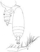 Issued from : W. Vervoort in Atlantide Report, 1965, 8. [p75., Fig.17]. As Scolecithricella (Amallothrix) marquesae. Female (from off Ghana): a, habitus (dorsal); b, posterior part cephalothorax and urosome (lateral right side); d, Mxp. Nota: Urosome 4-segmented; urosomal somites and caudal rami of proportional lengths 38:15:21:7:19 = 100.
|
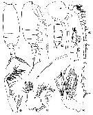
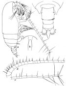 Issued from : W. Vervoort in Atlantide Report, 1965, 8. [p76., Fig.18]. As Scolecithricella (Amallothrix) marquesae. Female: a, habitus (lateral right side); b, rostrum (ventral); c, posterior part cephalothorax and urosome (dorsal); d-e, A1. Nota: Rotrum pointing downwards, with 2 fine filaments of considerable length that are hidden between the basal parts of the A1. Genital complex perfectly symmetrical in dorsal view; on the left side there is a short row of spinules; on the right side there are 2 of such rows. A1 24-segmented (segments 8 and 9 fused)
|
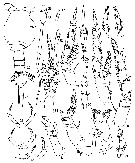
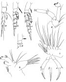 Issued from : W. Vervoort in Atlantide Report, 1965, 8. [p79., Fig.19]. As Scolecithricella (Amallothrix) marquesae. Female: a, P1; b, P2; c, P3; d, P4; e, P5; f, apex of terminal spine of P5; g, A2; h, Mx1; i, Mx2. Nota: Praecoxa of Mx1 with 7 strong, spiniform setae, the basal 3 of which are spinulose; the endites of coxa and basis, both carry 2 setae; basipodite with 3 internal setae; exopodite well developed but indistinctly separated from the basipodite, with 5 setae; endopodite unsegmented with 6 setae, 4 of which are apical; epipodite (coxal exite) with 7 setae ( As this description is based on the study of a single maxillule only the actual number may be slightly different).
|
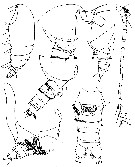 issued from : N.V. Vyshkvartzeva in Zoosyst. Ros., 2000 (2001), 9 (1). [p.81, Figs.1-7]. Female (from Atlantic): 1, habitus (lateral); 2, forehead (lateral); 3, thoracic segment 5 and urosome (lateral, right side); 4, idem with spermatophore; 5, thoracic segment 5 and urosome (lateral, left side); 6, urosome (ventral); 7, right A1.
|
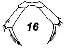
 issued from : N.V. Vyshkvartzeva in Zoosyst. Ros., 2000 (2001), 9 (1). [p.82, Figs.8-16]. Female: 8, thoracic segments 4-5 and urosome (dorsal); 9, forehead (dorsal); 10, A2; 11, Mx1; 12, Mx2; 13, Mxp; 14, P2; 15, P4; 16, P5.
|
 issued from : N.V. Vyshkvartzeva in Zoosyst. Ros., 2000 (2001), 9 (1). [p.84, Figs.17-38]. Female: 17, Md; 18, P1; 19, P2 (without exopodal segments 2-3); 20, P2 (without endopodal segments 2-3); 21, P2 endopod (dorsal view); 22, P2 endopod (ventral view); 23-25, P5. Male: 26, forehead (dorsal); 27, thoracic segment 5 and urosome (dorsal); 28, rostrum; 29, P2 (posterior surface); 30, P2 endopod (another specimen); 31, P2 (apical spine); 32, P3 (posterior surface); 33, P3 endopod (anterior surface); 34, P4; 35, P4 endopod (anterior surface); 36, P5; 37, 38, P5 (left distal segment and distal part of penultimate segment in different positions).
|
 issued from : N.V. Vyshkvartzeva in Zoosyst. Ros., 2000 (2001), 9 (1). [p.85, Figs.39-47]. Male: 39-40, habitus (dorsal and lateral, respectively); 41, forehead (lateral); ; 42, thoracic segment 5 with left leg of P5 and urosome (lateral, right side); 43, right A1; 44, Mx1; 45, Mx2; 46, Mxp; 47, P1.
|
 Issued from : V.N. Andronov in Russian Acad. Sci. P.P. Shirshov Inst. Oceanol. Atlantic Branch, Kaliningrad, 2014. [p.84, Fig.21: 16]. Scolecitrichopsis tenuipes after Vyshkvartzeva, 2001. Female P5.
| | | | | NZ: | 5 | | |
|
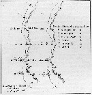
Distribution map of Scolecitrichopsis tenuipes by geographical zones
|
| | | | | |  issued from : U. Brenning in Wiss. Z. Wilhelm-Pieck-Univ. Rostock - 33. Jahrgang 1984. Mat.-nat. wiss. Reihe, 6. [p.6, Fig.2]. issued from : U. Brenning in Wiss. Z. Wilhelm-Pieck-Univ. Rostock - 33. Jahrgang 1984. Mat.-nat. wiss. Reihe, 6. [p.6, Fig.2].
Spatial distribution for Scolecithricella marquesae (= Scolecitrichopsis tenuipes) and other scolecithrids from 8° S - 26° N; 16°- 20° W, for different expeditions (V1: Dec. 1972- Jan. 1973; V2: Feb/Mar. 1973; VI: May 1974; IV: Jun./Jul. 1972). |
| | | | Loc: | | | off Angola, Congo, G. of Guinea, Ivorian shelf, off Ghana , off Mauritania, Red Sea, Sri Lanka, Indonesia-Malaysia (in A. Scott, 1909) | | | | N: | 11 | | | | Lg.: | | | (16) F: 1,24-1,152 ; (57) M: 1,4; (816) F: 1,48-1,30; M: 1,45-1,38; (1111) F: 1,34-1,44; {F: 1,152-1,480; M: 1,380-1,450} | | | | Rem.: | epi-mesopelagic.
For Vervoort (1965, p.80) the specimens described above are identical with the species mentioned by Marques (1959, p.16) as Amallothrix sp. from the coastal waters of Angola. There are very small differences in the spinulation of the legs, that may be due to individual variation, whilst the fact that Marques figured the endopodite of P2 as being 3-segmented may be due to abnormality in her specimen, as in the holotype the endopodite of P3 on the right side also shows abnormal structure. | | | Last update : 30/06/2016 | |
|
|
 Any use of this site for a publication will be mentioned with the following reference : Any use of this site for a publication will be mentioned with the following reference :
Razouls C., Desreumaux N., Kouwenberg J. and de Bovée F., 2005-2026. - Biodiversity of Marine Planktonic Copepods (morphology, geographical distribution and biological data). Sorbonne University, CNRS. Available at http://copepodes.obs-banyuls.fr/en [Accessed March 21, 2026] © copyright 2005-2026 Sorbonne University, CNRS
|
|
|
|


;)
;)
;)
;)
;)
;)
;)
;)

;)
