|
|
 |
Fiche d'espèce de Copépode |
|
|
Calanoida ( Ordre ) |
|
|
|
Clausocalanoidea ( Superfamille ) |
|
|
|
Scolecitrichidae ( Famille ) |
|
|
|
Scolecitrichopsis ( Genre ) |
|
|
| |
Scolecitrichopsis tenuipes (T. Scott, 1894) (F,M) | |
| | | | | | | Syn.: | Scolecithrix tenuipes T. Scott, 1894 b (p.48, Descr.M, figs.M); Giesbrecht & Schmeil, 1898 (p.47, Rem.M); Thompson & Scott, 1903 (p.234, 245); Bradford & al., 1983 (p.104);
Scolecithricella tenuipes : A. Scott, 1909 (p.92, Rem.M); Sewell, 1948 (p.516, 517, 523, 546); Bainbridge, 1972 (p.61, Appendix Table I: vertical distribution vs day/night); Dessier, 1979 (p.205);
Scolecithricella (Amallothrix) marquesae Vervoort, 1965 (p.74, Descr.F, figs.F, Rem.);
Amallothrix sp. Marques, 1958 (p.212);
Amallothrix marquesae : Vives, 1982 (p.292);
Scolecithricella (Amallothrix) marquesae : Vervoort, 1965 (p.79, figs.F);
Scolecithricella marquesae : Binet & al., 1972 (p.68); Bradford, 1973 (p.146); Bradford & al., 1983 (p.78, 104, Rem.); Brenning, 1984 (p.5, Rem.); 1985 a (p.16, 28, Table 2); Vives & Shmeleva, 2007 (p.794, figs.F, Rem.) | | | | Ref.: | | | Vyshkvartzeva, 1999 (2000) (p.221, Rem.); 2001 (p.83, Redescr., figs.F,M, Rem.) | 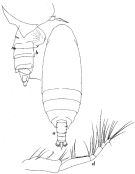 Issued from : W. Vervoort in Atlantide Report, 1965, 8. [p75., Fig.17]. As Scolecithricella (Amallothrix) marquesae. Female (from off Ghana): a, habitus (dorsal); b, posterior part cephalothorax and urosome (lateral right side); d, Mxp. Nota: Urosome 4-segmented; urosomal somites and caudal rami of proportional lengths 38:15:21:7:19 = 100.
|
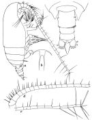 Issued from : W. Vervoort in Atlantide Report, 1965, 8. [p76., Fig.18]. As Scolecithricella (Amallothrix) marquesae. Female: a, habitus (lateral right side); b, rostrum (ventral); c, posterior part cephalothorax and urosome (dorsal); d-e, A1. Nota: Rotrum pointing downwards, with 2 fine filaments of considerable length that are hidden between the basal parts of the A1. Genital complex perfectly symmetrical in dorsal view; on the left side there is a short row of spinules; on the right side there are 2 of such rows. A1 24-segmented (segments 8 and 9 fused)
|
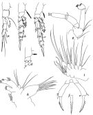 Issued from : W. Vervoort in Atlantide Report, 1965, 8. [p79., Fig.19]. As Scolecithricella (Amallothrix) marquesae. Female: a, P1; b, P2; c, P3; d, P4; e, P5; f, apex of terminal spine of P5; g, A2; h, Mx1; i, Mx2. Nota: Praecoxa of Mx1 with 7 strong, spiniform setae, the basal 3 of which are spinulose; the endites of coxa and basis, both carry 2 setae; basipodite with 3 internal setae; exopodite well developed but indistinctly separated from the basipodite, with 5 setae; endopodite unsegmented with 6 setae, 4 of which are apical; epipodite (coxal exite) with 7 setae ( As this description is based on the study of a single maxillule only the actual number may be slightly different).
|
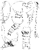 issued from : N.V. Vyshkvartzeva in Zoosyst. Ros., 2000 (2001), 9 (1). [p.81, Figs.1-7]. Female (from Atlantic): 1, habitus (lateral); 2, forehead (lateral); 3, thoracic segment 5 and urosome (lateral, right side); 4, idem with spermatophore; 5, thoracic segment 5 and urosome (lateral, left side); 6, urosome (ventral); 7, right A1.
|
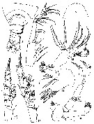 issued from : N.V. Vyshkvartzeva in Zoosyst. Ros., 2000 (2001), 9 (1). [p.82, Figs.8-16]. Female: 8, thoracic segments 4-5 and urosome (dorsal); 9, forehead (dorsal); 10, A2; 11, Mx1; 12, Mx2; 13, Mxp; 14, P2; 15, P4; 16, P5.
|
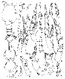 issued from : N.V. Vyshkvartzeva in Zoosyst. Ros., 2000 (2001), 9 (1). [p.84, Figs.17-38]. Female: 17, Md; 18, P1; 19, P2 (without exopodal segments 2-3); 20, P2 (without endopodal segments 2-3); 21, P2 endopod (dorsal view); 22, P2 endopod (ventral view); 23-25, P5. Male: 26, forehead (dorsal); 27, thoracic segment 5 and urosome (dorsal); 28, rostrum; 29, P2 (posterior surface); 30, P2 endopod (another specimen); 31, P2 (apical spine); 32, P3 (posterior surface); 33, P3 endopod (anterior surface); 34, P4; 35, P4 endopod (anterior surface); 36, P5; 37, 38, P5 (left distal segment and distal part of penultimate segment in different positions).
|
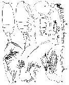 issued from : N.V. Vyshkvartzeva in Zoosyst. Ros., 2000 (2001), 9 (1). [p.85, Figs.39-47]. Male: 39-40, habitus (dorsal and lateral, respectively); 41, forehead (lateral); ; 42, thoracic segment 5 with left leg of P5 and urosome (lateral, right side); 43, right A1; 44, Mx1; 45, Mx2; 46, Mxp; 47, P1.
|
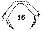 Issued from : V.N. Andronov in Russian Acad. Sci. P.P. Shirshov Inst. Oceanol. Atlantic Branch, Kaliningrad, 2014. [p.84, Fig.21: 16]. Scolecitrichopsis tenuipes after Vyshkvartzeva, 2001. Female P5.
| | | | | NZ: | 5 | | |
|
Carte de distribution de Scolecitrichopsis tenuipes par zones géographiques
|
| | | | | | 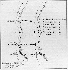 issued from : U. Brenning in Wiss. Z. Wilhelm-Pieck-Univ. Rostock - 33. Jahrgang 1984. Mat.-nat. wiss. Reihe, 6. [p.6, Fig.2]. issued from : U. Brenning in Wiss. Z. Wilhelm-Pieck-Univ. Rostock - 33. Jahrgang 1984. Mat.-nat. wiss. Reihe, 6. [p.6, Fig.2].
Spatial distribution for Scolecithricella marquesae (= Scolecitrichopsis tenuipes) and other scolecithrids from 8° S - 26° N; 16°- 20° W, for different expeditions (V1: Dec. 1972- Jan. 1973; V2: Feb/Mar. 1973; VI: May 1974; IV: Jun./Jul. 1972). |
| | | | Loc: | | | off Angola, Congo, G. of Guinea, Ivorian shelf, off Ghana , off Mauritania, Red Sea, Sri Lanka, Indonesia-Malaysia (in A. Scott, 1909) | | | | N: | 11 | | | | Lg.: | | | (16) F: 1,24-1,152 ; (57) M: 1,4; (816) F: 1,48-1,30; M: 1,45-1,38; (1111) F: 1,34-1,44; {F: 1,152-1,480; M: 1,380-1,450} | | | | Rem.: | épi-mésopélagique.
Voir aussi les remarques en anglais | | | Dernière mise à jour : 30/06/2016 | |
|
|
 Toute utilisation de ce site pour une publication sera mentionnée avec la référence suivante : Toute utilisation de ce site pour une publication sera mentionnée avec la référence suivante :
Razouls C., Desreumaux N., Kouwenberg J. et de Bovée F., 2005-2026. - Biodiversité des Copépodes planctoniques marins (morphologie, répartition géographique et données biologiques). Sorbonne Université, CNRS. Disponible sur http://copepodes.obs-banyuls.fr [Accédé le 22 juin 2026] © copyright 2005-2026 Sorbonne Université, CNRS
|
|
|
|




;)
;)
;)
;)
;)
;)
;)
;)

;)