|
|
 |
|
Calanoida ( Order ) |
|
|
|
Clausocalanoidea ( Superfamily ) |
|
|
|
Stephidae ( Family ) |
|
|
|
Stephos ( Genus ) |
|
|
| |
Stephos cryptospinosus Zagami, Campolmi & Costanzo, 2000 (F,M) | |
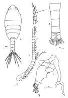
| | | | | | | Ref.: | | | Zagami & al., 2000 (p.16, figs.F,M); Vives & Shmeleva, 2007 (p.850, figs.F,M, Rem.); Brylinski & Courtot, 2019 (p.3, Rem.: F, M, figs.F, M) | 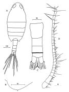 issued from : G. Zagami, M. Campolmi & G. Costanzo in Journal Plankton Res., 2000, 22 (1). [p.17, Fig.1] Female: A, habitus (dorsal); B, posterolateral margin of 5th pedigerous somite; C, urosome (dorsal); D, A1. All scales in microns.
|
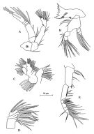 issued from : G. Zagami, M. Campolmi & G. Costanzo in Journal Plankton Res., 2000, 22 (1). [p.18, Fig.2]. Female: A, A2; B, Md; C, Mx1; D, Mx2; E, Mxp.
|
 issued from : G. Zagami, M. Campolmi & G. Costanzo in Journal Plankton Res., 2000, 22 (1). [p.20]. Female: A, P1; B, P2; C, P3; D, P4; E, P5.
|
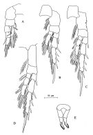
 issued from : G. Zagami, M. Campolmi & G. Costanzo in Journal Plankton Res., 2000, 22 (1). [p.22, Fig.4]. Male: A, habitus (dorsal); B, urosome (dorsal); C, A1; D, P5. All scales in microns.
|
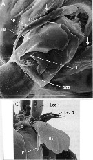 Issued from : J.-M. Brylinski & L. Courcot in Bioinvasions Records, 2019, 8. [p.4, Fig.2, A, C]. Female (from 47°52'06''N, 03°55'00''W): A, urosome (ventral view); C, same (lateral view ). Scale bar: 100 µm. Sp: spermatophore; DGS: genital double somite; HS: hyaline sheath; L: lappets of genital operculum; ; P: point of the hyaline sheath. Arrows: ''cryptospine'' on last prosomite. Nota: - Posterior prosomal margins slightly asymmetrical. Minute spinule present dorso-laterally on each side (demonstrated using SEM observations). - Urosome 4-segmented. - Genital double-somite symmetrical; central genital aperture framed by 2 asymmetrical and very fine lappets (only observable with SEM). - Tubular spematothore implanted directly in genital opening, attached by long tubule. - When carrying a spermatophore, the genital double-somite is partially covered by a hyaline sheath which tapers to a point dorsally; this structure appeared fixed to the right side of the genital double-somite and extends forward as far as the basal articulation of the right P4.
|
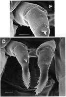 Issued from : J.-M. Brylinski & L. Courcot in Bioinvasions Records, 2019, 8. [p.4, Fig.2, E, D]. Female: D-E, P5. Scale bar: 10 µm. Arrows: patch of micro-spinules. Nota: - P5 uniramous, 2-segmented; proximal segment naked, distal segment armed with 1 short central spine on middle of anterior surface; patch of very small spinules present proximally to the spine. Apical part of P5 slightly asymmetrical with row of 8-9 or 12-15 spinules on outer margin , and 6-7 or 4-5 spinules on inner margin, of right and left legs, respectively.
|
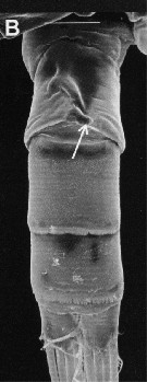 Issued from : J.-M. Brylinski & L. Courcot in Bioinvasions Records, 2019, 8. [p.45 Fig.3, B]. Male: B, urosome (ventral view). Scale: 40 µm. Arrow: short process directed towards left side on the 2nd urosomite. Nota: - Posterior prosomal margin symmetrical with minute spinule (as in female). - Urosome 5-segmented. - P5 elongate and markedly asymmetrical, both legs uniramous. Right bleg slender, 4-segmented; 1st and 2nd segments short, unarmed; 3rd segment elonjgate bearing acute outer process near base, slightly curving inwards; 4th segment comprising 2 processes of unequal length. left leg 5-segmented, 1st to 3rd segments short, unarmed, 4th segment rpbust and swollen; 5th segment complex with 2 pairs of long,0 wide lamellar spines, 1 pair of long, narrow lamellar spines, and 1 row of 8 wide, amber-coloured claws directed posteriorly.
| | | | | Compl. Ref.: | | | Mazzocchi & Di Capua, 2010 (p.427); Belmonte, 2018 (p.273, Table I: Italian zones) | | | | NZ: | 2 | | |
|
Distribution map of Stephos cryptospinosus by geographical zones
|
| | | | | | 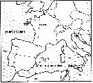 Issued from : J.-M. Brylinski & L. Courcot in Bioinvasions Rec., 2019, 8 (2). [p.358, Fig.1] Issued from : J.-M. Brylinski & L. Courcot in Bioinvasions Rec., 2019, 8 (2). [p.358, Fig.1]
Global distribution of Stephos cryptospinus (black star) and S. marsalensis (white star). C : Concarneau ; W: Wimereux . |
| | | | Loc: | | | W Medit. (W Sicily: Stagnone di Marsala); NE Atlantic (Concarneau) | | | | N: | 4 | | | | Lg.: | | | (838) F: 0,86; M: 0,78; (1226) F: 0,90; M; 0,75, 0,85; {F: 0,86-0,90; M: 0,75-0,85} | | | | Rem.: | coastal (hyperbenthic, 2-3 m).
The species belongs to ''group III'' (see genus Stephos) in which segment 4 of the left male P5 is swollen, according to the classification of Bradford-Grieve (1999).
For Brylinski & Courcot (2019, p.362) this species named cryptospinus because it had ''a small spinous process on the posterolateral margin of the last prosomite'', but not visible using light microscopy because of its small size (4 µm), only visible using SEM , but after Zagami, the spinous process is visible. Some morphological differences were noted with regard to the original description and some supplementary observations are provided with SEM.
The genital aperture of female was described as ''closed off by a single unarmed operculum" (Zagami & al., 2000: fig.5) but in the Concarneau's material the operculum comprises 2 asymmetrical lappets (not visible using microscopy because too fine and transparent).; The original drawing of P5 female (Zagami & al., 2000) was inverted because the central spine (s) on the distal segment is normally located on the anterior surface in all Stephos sp. The inner margin bears fewer spinules on the left than on the right leg; both Concarneau's data and Sicilian data showed variability in the number of these marginal spinules on P5. The right leg is slightly larger than left one and twisted towards the left.
Concerning the left P5 male from Zagami & al. (2000, fig.4D), the 2 rounded inner margin processes on the 4th segment, with the depression between these 2 processes is a artefact, probably resulting from shrinkage of the internal tissues within the cuticle. Therefore, S. cryptospinosus belongs to type III, in which segment 4 of the left male P5 is swollen according to the classification of Bradford-Grieve (1999).
Brylinski & Coucot (2019, p.364-365) summarize the occurrence of the ''hyaline sheath' in various species of calanoid copepods. For example such structure was observed in S. cryptospinosus, but the authors suspect that it is not a general feature of the genus, not found in S. scotti. Bradford-Grieve (1999) noted that the female of Stephos, "when carrying a spermatophore, the genital double-somite is covered by a hyaline sheath of a complex structure". The origin of this sheath is uncertain but Fosshagen (1870) suggested that "it seems to be secreted from 2 gland-like structures, one on either side of the last prosomal segment next to the genital segment"., but the authors did not find these glandular pores in either sex on any species studied using SEM. The hyaline sheath differs in shape in each species butb the precise position can vary from specimen to specimen within a species (Fosshagen, 1970), as also shownj in the Concarneau's specimens.. Observations of the authors suggest it is producede by the male at the same time as the spermatophore. The variable positioning on the female might reflect variation in copulatory behaviour of males.
Most calanoid copepods produce simple tubular spermatophores as in the genus Stephos, but several produce more complex spermatophores, for example, some species of the subgenus Tortanus (Atortus) have highly complex coupling device (Barthélémy & al., 2003). In Centropages sp., the tubular spermatophore is connected via a bipartite coupling device (Lee, 1972).
The authors hypothesis that the hyaline sheath in Stephos is a relict structure derived from an ancestral complex spermatophore.After Ohtsuka & Huys (2001), such structures can prevent subsequent mating by other males, but the efficiency of such system is uncertain for Brylinski & Courcot. | | | Last update : 19/12/2020 | |
|
|
 Any use of this site for a publication will be mentioned with the following reference : Any use of this site for a publication will be mentioned with the following reference :
Razouls C., Desreumaux N., Kouwenberg J. and de Bovée F., 2005-2026. - Biodiversity of Marine Planktonic Copepods (morphology, geographical distribution and biological data). Sorbonne University, CNRS. Available at http://copepodes.obs-banyuls.fr/en [Accessed March 23, 2026] © copyright 2005-2026 Sorbonne University, CNRS
|
|
|
|


;)
;)
;)
;)
;)
;)
;)

;)
