|
|
 |
Fiche d'espèce de Copépode |
|
|
Calanoida ( Ordre ) |
|
|
|
Clausocalanoidea ( Superfamille ) |
|
|
|
Stephidae ( Famille ) |
|
|
|
Stephos ( Genre ) |
|
|
| |
Stephos cryptospinosus Zagami, Campolmi & Costanzo, 2000 (F,M) | |
| | | | | | | Ref.: | | | Zagami & al., 2000 (p.16, figs.F,M); Vives & Shmeleva, 2007 (p.850, figs.F,M, Rem.); Brylinski & Courtot, 2019 (p.3, Rem.: F, M, figs.F, M) | 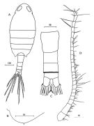 issued from : G. Zagami, M. Campolmi & G. Costanzo in Journal Plankton Res., 2000, 22 (1). [p.17, Fig.1] Female: A, habitus (dorsal); B, posterolateral margin of 5th pedigerous somite; C, urosome (dorsal); D, A1. All scales in microns.
|
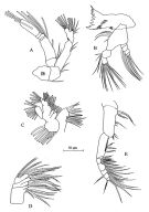 issued from : G. Zagami, M. Campolmi & G. Costanzo in Journal Plankton Res., 2000, 22 (1). [p.18, Fig.2]. Female: A, A2; B, Md; C, Mx1; D, Mx2; E, Mxp.
|
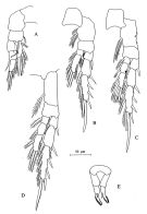 issued from : G. Zagami, M. Campolmi & G. Costanzo in Journal Plankton Res., 2000, 22 (1). [p.20]. Female: A, P1; B, P2; C, P3; D, P4; E, P5.
|
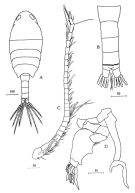 issued from : G. Zagami, M. Campolmi & G. Costanzo in Journal Plankton Res., 2000, 22 (1). [p.22, Fig.4]. Male: A, habitus (dorsal); B, urosome (dorsal); C, A1; D, P5. All scales in microns.
|
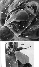 Issued from : J.-M. Brylinski & L. Courcot in Bioinvasions Records, 2019, 8. [p.4, Fig.2, A, C]. Female (from 47°52'06''N, 03°55'00''W): A, urosome (ventral view); C, same (lateral view ). Scale bar: 100 µm. Sp: spermatophore; DGS: genital double somite; HS: hyaline sheath; L: lappets of genital operculum; ; P: point of the hyaline sheath. Arrows: ''cryptospine'' on last prosomite. Nota: - Posterior prosomal margins slightly asymmetrical. Minute spinule present dorso-laterally on each side (demonstrated using SEM observations). - Urosome 4-segmented. - Genital double-somite symmetrical; central genital aperture framed by 2 asymmetrical and very fine lappets (only observable with SEM). - Tubular spematothore implanted directly in genital opening, attached by long tubule. - When carrying a spermatophore, the genital double-somite is partially covered by a hyaline sheath which tapers to a point dorsally; this structure appeared fixed to the right side of the genital double-somite and extends forward as far as the basal articulation of the right P4.
|
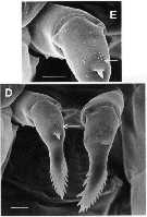 Issued from : J.-M. Brylinski & L. Courcot in Bioinvasions Records, 2019, 8. [p.4, Fig.2, E, D]. Female: D-E, P5. Scale bar: 10 µm. Arrows: patch of micro-spinules. Nota: - P5 uniramous, 2-segmented; proximal segment naked, distal segment armed with 1 short central spine on middle of anterior surface; patch of very small spinules present proximally to the spine. Apical part of P5 slightly asymmetrical with row of 8-9 or 12-15 spinules on outer margin , and 6-7 or 4-5 spinules on inner margin, of right and left legs, respectively.
|
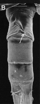 Issued from : J.-M. Brylinski & L. Courcot in Bioinvasions Records, 2019, 8. [p.45 Fig.3, B]. Male: B, urosome (ventral view). Scale: 40 µm. Arrow: short process directed towards left side on the 2nd urosomite. Nota: - Posterior prosomal margin symmetrical with minute spinule (as in female). - Urosome 5-segmented. - P5 elongate and markedly asymmetrical, both legs uniramous. Right bleg slender, 4-segmented; 1st and 2nd segments short, unarmed; 3rd segment elonjgate bearing acute outer process near base, slightly curving inwards; 4th segment comprising 2 processes of unequal length. left leg 5-segmented, 1st to 3rd segments short, unarmed, 4th segment rpbust and swollen; 5th segment complex with 2 pairs of long,0 wide lamellar spines, 1 pair of long, narrow lamellar spines, and 1 row of 8 wide, amber-coloured claws directed posteriorly.
| | | | | Ref. compl.: | | | Mazzocchi & Di Capua, 2010 (p.427); Belmonte, 2018 (p.273, Table I: Italian zones) | | | | NZ: | 2 | | |
|
Carte de distribution de Stephos cryptospinosus par zones géographiques
|
| | | | | | 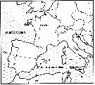 Issued from : J.-M. Brylinski & L. Courcot in Bioinvasions Rec., 2019, 8 (2). [p.358, Fig.1] Issued from : J.-M. Brylinski & L. Courcot in Bioinvasions Rec., 2019, 8 (2). [p.358, Fig.1]
Global distribution of Stephos cryptospinus (black star) and S. marsalensis (white star). C : Concarneau ; W: Wimereux . |
| | | | Loc: | | | W Medit. (W Sicily: Stagnone di Marsala); NE Atlantic (Concarneau) | | | | N: | 4 | | | | Lg.: | | | (838) F: 0,86; M: 0,78; (1226) F: 0,90; M; 0,75, 0,85; {F: 0,86-0,90; M: 0,75-0,85} | | | | Rem.: | côtière (hyperbentique, 2-3 m).
Voir aussi les remarques en anglais | | | Dernière mise à jour : 19/12/2020 | |
|
|
 Toute utilisation de ce site pour une publication sera mentionnée avec la référence suivante : Toute utilisation de ce site pour une publication sera mentionnée avec la référence suivante :
Razouls C., Desreumaux N., Kouwenberg J. et de Bovée F., 2005-2026. - Biodiversité des Copépodes planctoniques marins (morphologie, répartition géographique et données biologiques). Sorbonne Université, CNRS. Disponible sur http://copepodes.obs-banyuls.fr [Accédé le 08 janvier 2026] © copyright 2005-2026 Sorbonne Université, CNRS
|
|
|
|




;)
;)
;)
;)
;)
;)
;)

;)