|
|
 |
|
Monstrilloida ( Order ) |
|
|
|
Monstrillidae ( Family ) |
|
|
|
Monstrillopsis ( Genus ) |
|
|
| |
Monstrillopsis dubioides Suarez-Morales, 2004 (F,M) | |
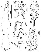
| | | | | | | Syn.: | Monstrillopsis dubia : Sars, 1921 (p.26, figs.F,M); Rose, 1933 a (p.352, figs.F,M); Vilela, 1968 (p.45, fig.F); Razouls, 1972 (Annexe: p.155); 1973 (p.467); Huys & Boxshall, 1991 (p.168: figs.M); | | | | Ref.: | | | in Suarez-Morales & Ivanenko, 2004 (p.42, figs.F, Rem.); Suarez-Morales & al., 2006 (p.101, 105, Tab.1); Vives & Shmeleva, 2010 (p.188, figs.F, Rem.); Suarez-Morales, 2011 (p.10) |  issued from : E. Suarez-Morales & V.V. Ivanenko in Arctic, 2004, 57 (1). [p.43, Fig.8]. Female (from outside Trondheimsfjord, Norway): A, cephalothorax (lateral); B, 5th pedigerous somite and genital double-somite (lateral); C, idem (ventral); D, 2nd, 3rd and 4th pedigerous somites (4th separated; lateral); E, last part of urosome and caudal rami.
|
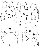
 issued from : E. Suarez-Morales & V.V. Ivanenko in Arctic, 2004, 57 (1). [p.43, Fig.9]. Female: A, cephalic area with part of A1 (dorsal); B, exopod of P1; C, two distal segments of exopod of P2, P3 or P4; D, urosome (lateral, another specimen); E, cephalic area (dorsal, from second specimen); F, idem (ventral; from the first specimen).
|
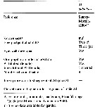
 issued from : E. Suarez-Morales & V.V. Ivanenko in Arctic, 2004, 57 (1). [p.42, Table 1]. Comparison of taxonomic features and morphometry in females of selected species of Monstrillopsis. A1 = antennule; AN = anal somite; CT = cephalothorax; FP = 1st pediger; GS = genital double-somite; LA = last antennular segment; TL = total length; UR = urosome. All ratios are presented in percentage form.
|
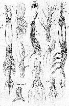
 issued from : G.O. Sars in An Account of the Crustacea of Norway, with short descriptions and figures of all the species, 1921 a, 8. [Pl. XIV]. As Monstrillopsis dubia. Female and Male (from Bejan: outside of Trondhjem Fjord). Female: Nota: Cephalic segment exceeding the remaining part of the body by 1/3 of its length. Eye very conspicuous, with dark pigment and all 3 lenses well developed. A1 4-segmented, exceeding somewhat in length 1/3 of the cephalic segment; the last segment is fully as long as the other 3 combined, none of the setae ramified. Oral tubule well marked, located near the frontal part of the head. natatory legs with the exopod considerably longer than endopod; P5 rather narrow at the base, but considerably widdening towards the end, which is produced to a conical lappet, across the base of which 3 slender setae are attached, inner edge of the leg produced to a similar lappet, which is quite smooth. Genital segment a little longer than the other 2 segments combined. Ovigerous spines of moderate length. Anal segment somewhat flattened and sharply defined from the rather small middle segment. Caudal rami exceeding somewhat in length the 2 preceeding segments combined, slightly divergent, each ramus with 4 setae, 1 about in the middle of the outer edge, 2 at the apex, and 1 inside at some distance from the end. Male: Nota: Cephalic segment somewhat club-shaped, and scarcely exceeding half the length of the body. Eye more developed than in female, with the ventral lens prominent and highly refractive. A1 5-segmented, considerably exceeding half the length of the cephalic segment; the last segment very mobile segment abruptly attenuated distally. Abdomen 4-segmented, the first of which is produced below to a rather large copulative appendage divided at the end into 2 diverging subcylindral rami.
|
 Issued from : E. Suarez-Morales, A. Bello-Smith & S. Palma in Zool. Anz., 2006, 245. [p.103, Table 1]. Structural characteristics. Morphological characters for comparative morphology of the species assigned to the genus Monstrillopsis by different authorities. See : dubia, M. fosshageni, M. reticulata, M. sarsi, Cymbasoma gracilis, M. ferrari, M. zernovi, M. angustipes, Monstrillopsis sp. Key for the identification of the currently known species of Monstrillopsis (female only) after Suarez-Morales & al. (2006, p.105): 1 - Outer (exopodal) lobe of P5 with distal elongation; last antennular segment relatively long, more than 50% of A1 length; large species (more than 3 mm) ..... 2. 2 - Genital doubble-somite relatively long, representing more than 55% of urosome length; A1 length about or less than 20% of total bopdy length, element 1 (on 1st segment) reduced; caudal rami elongate 2.2 times longer than wide.
| | | | | Compl. Ref.: | | | ? Oliveira Dias, 1996 (p.253); ? Bernier & al., 2002 (p.651, tab.1); Suarez-Morales, 2011 (p.12) | | | | NZ: | 2 + 2 doubtful | | |
|
Distribution map of Monstrillopsis dubioides by geographical zones
|
| | | | | | | Loc: | | | Norway (Bejan: outside the Trondheim Fjord), Portugal, ? Medit. (North Africa, Banyuls), ? Brazil | | | | N: | 5 ? | | | | Lg.: | | | (307) F: 3,8; M: 2,1; (327) F: 2,3; ? (466) F: 2,48-1,19; {F: 2,3-3,8; M: 2,1} | | | | Rem.: | The locality records in the Mediterranean and from Brazil are to be confirmed.
Tis species is close to M. ferrarii and M. chilensis.
For Suarez-Morales & Ivanenko (2004, p.43) several characters present in Monstrillopsis dubia Scott, 1904, differ from those described by Sars (1921) for supposedly the same nominal species. The main differences between these two species reside in the proportions of the body parts, particularly in the urosome; the genital double somite, the 5th pedigerous somite, and the anal somite differ in shape and relative size in the two species (table 1); an additional difference is the relative length of A1, which are very long in M. dubia (sensu Scott, 1904), 32.5 % of total body length, and shorter (22 %) in M. dubioides.
After Lee J. & al; (2016, p.421), genitalia male with a well developed, long median shaft and relatively short, ovoidal or globose lateral lappets that are more or less separated from the median shaft belongs subgroup ''Type I'' (Suarez-Morales & McKinnon, 2014).
See remarks in Suarez-Morales & al. (2006, p.101) concerning comparison with M. chilensis. | | | Last update : 23/08/2021 | |
|
|
 Any use of this site for a publication will be mentioned with the following reference : Any use of this site for a publication will be mentioned with the following reference :
Razouls C., Desreumaux N., Kouwenberg J. and de Bovée F., 2005-2024. - Biodiversity of Marine Planktonic Copepods (morphology, geographical distribution and biological data). Sorbonne University, CNRS. Available at http://copepodes.obs-banyuls.fr/en [Accessed November 21, 2024] © copyright 2005-2024 Sorbonne University, CNRS
|
|
 |
 |


