|
|
 |
Fiche d'espèce de Copépode |
|
|
Monstrilloida ( Ordre ) |
|
|
|
Monstrillidae ( Famille ) |
|
|
|
Monstrillopsis ( Genre ) |
|
|
| |
Monstrillopsis dubioides Suarez-Morales, 2004 (F,M) | |
| | | | | | | Syn.: | Monstrillopsis dubia : Sars, 1921 (p.26, figs.F,M); Rose, 1933 a (p.352, figs.F,M); Vilela, 1968 (p.45, fig.F); Razouls, 1972 (Annexe: p.155); 1973 (p.467); Huys & Boxshall, 1991 (p.168: figs.M); | | | | Ref.: | | | in Suarez-Morales & Ivanenko, 2004 (p.42, figs.F, Rem.); Suarez-Morales & al., 2006 (p.101, 105, Tab.1); Vives & Shmeleva, 2010 (p.188, figs.F, Rem.); Suarez-Morales, 2011 (p.10) | 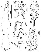 issued from : E. Suarez-Morales & V.V. Ivanenko in Arctic, 2004, 57 (1). [p.43, Fig.8]. Female (from outside Trondheimsfjord, Norway): A, cephalothorax (lateral); B, 5th pedigerous somite and genital double-somite (lateral); C, idem (ventral); D, 2nd, 3rd and 4th pedigerous somites (4th separated; lateral); E, last part of urosome and caudal rami.
|
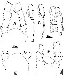 issued from : E. Suarez-Morales & V.V. Ivanenko in Arctic, 2004, 57 (1). [p.43, Fig.9]. Female: A, cephalic area with part of A1 (dorsal); B, exopod of P1; C, two distal segments of exopod of P2, P3 or P4; D, urosome (lateral, another specimen); E, cephalic area (dorsal, from second specimen); F, idem (ventral; from the first specimen).
|
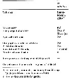
 issued from : E. Suarez-Morales & V.V. Ivanenko in Arctic, 2004, 57 (1). [p.42, Table 1]. Comparison of taxonomic features and morphometry in females of selected species of Monstrillopsis. A1 = antennule; AN = anal somite; CT = cephalothorax; FP = 1st pediger; GS = genital double-somite; LA = last antennular segment; TL = total length; UR = urosome. All ratios are presented in percentage form.
|
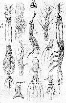 issued from : G.O. Sars in An Account of the Crustacea of Norway, with short descriptions and figures of all the species, 1921 a, 8. [Pl. XIV]. As Monstrillopsis dubia. Female and Male (from Bejan: outside of Trondhjem Fjord). Female: Nota: Cephalic segment exceeding the remaining part of the body by 1/3 of its length. Eye very conspicuous, with dark pigment and all 3 lenses well developed. A1 4-segmented, exceeding somewhat in length 1/3 of the cephalic segment; the last segment is fully as long as the other 3 combined, none of the setae ramified. Oral tubule well marked, located near the frontal part of the head. natatory legs with the exopod considerably longer than endopod; P5 rather narrow at the base, but considerably widdening towards the end, which is produced to a conical lappet, across the base of which 3 slender setae are attached, inner edge of the leg produced to a similar lappet, which is quite smooth. Genital segment a little longer than the other 2 segments combined. Ovigerous spines of moderate length. Anal segment somewhat flattened and sharply defined from the rather small middle segment. Caudal rami exceeding somewhat in length the 2 preceeding segments combined, slightly divergent, each ramus with 4 setae, 1 about in the middle of the outer edge, 2 at the apex, and 1 inside at some distance from the end. Male: Nota: Cephalic segment somewhat club-shaped, and scarcely exceeding half the length of the body. Eye more developed than in female, with the ventral lens prominent and highly refractive. A1 5-segmented, considerably exceeding half the length of the cephalic segment; the last segment very mobile segment abruptly attenuated distally. Abdomen 4-segmented, the first of which is produced below to a rather large copulative appendage divided at the end into 2 diverging subcylindral rami.
|
 Issued from : E. Suarez-Morales, A. Bello-Smith & S. Palma in Zool. Anz., 2006, 245. [p.103, Table 1]. Structural characteristics. Morphological characters for comparative morphology of the species assigned to the genus Monstrillopsis by different authorities. See : dubia, M. fosshageni, M. reticulata, M. sarsi, Cymbasoma gracilis, M. ferrari, M. zernovi, M. angustipes, Monstrillopsis sp. Key for the identification of the currently known species of Monstrillopsis (female only) after Suarez-Morales & al. (2006, p.105): 1 - Outer (exopodal) lobe of P5 with distal elongation; last antennular segment relatively long, more than 50% of A1 length; large species (more than 3 mm) ..... 2. 2 - Genital doubble-somite relatively long, representing more than 55% of urosome length; A1 length about or less than 20% of total bopdy length, element 1 (on 1st segment) reduced; caudal rami elongate 2.2 times longer than wide.
| | | | | Ref. compl.: | | | ? Oliveira Dias, 1996 (p.253); ? Bernier & al., 2002 (p.651, tab.1); Suarez-Morales, 2011 (p.12) | | | | NZ: | 2 + 2 douteuses | | |
|
Carte de distribution de Monstrillopsis dubioides par zones géographiques
|
| | | | | | | Loc: | | | Norway (Bejan: outside the Trondheim Fjord), Portugal, ? Medit. (North Africa, Banyuls), ? Brazil | | | | N: | 5 ? | | | | Lg.: | | | (307) F: 3,8; M: 2,1; (327) F: 2,3; ? (466) F: 2,48-1,19; {F: 2,3-3,8; M: 2,1} | | | | Rem.: | Les localisations en Méditerranée et au Brésil sont à confirmer.
Voir aussi les remarques en anglais | | | Dernière mise à jour : 23/08/2021 | |
|
|
 Toute utilisation de ce site pour une publication sera mentionnée avec la référence suivante : Toute utilisation de ce site pour une publication sera mentionnée avec la référence suivante :
Razouls C., Desreumaux N., Kouwenberg J. et de Bovée F., 2005-2025. - Biodiversité des Copépodes planctoniques marins (morphologie, répartition géographique et données biologiques). Sorbonne Université, CNRS. Disponible sur http://copepodes.obs-banyuls.fr [Accédé le 03 décembre 2025] © copyright 2005-2025 Sorbonne Université, CNRS
|
|
|
|




;)
;)
;)
;)
;)