|
|
 |
|
Harpacticoida ( Order ) |
|
|
|
Cervinioidea ( Superfamily ) |
|
|
|
Aegisthidae ( Family ) |
|
|
|
Scabrantenna ( Genus ) |
|
|
| |
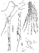
Scabrantenna yooi Lee & Huys, 2000 (F,M) | |
| | | | | | | Ref.: | | | Lee & Huys, 2000 (p.27, figs.F,M) | 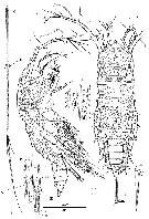 issued from : W. Lee & R. Huys in Zool. J. Linnean Soc., 2000, 129. [p.28, Fig.16]. Female (from Okinawa Trough): A, habitus (dorsal (distal 4/5 of caudal rami omitted); B, idem (lateral); C, caudal rami distal 4/5 (dorsal); D, ventral pore corresponding to original position of seta I (arrowed in A); E, seta II (dorsal); F, distal end of rami showing insertion sites of setae II-VII (dorsal).. Scales in microns. Nota: Prosome with complex surface reticulation consisting of anastomosing pattern of longutudinal and transversal lamellae; additional ornamentation consisting of sensillae and pores, paricularly around posterior margin of somites (Pediger 1 somite without sensillae, conspicuous aggregation of paired pores present middorsally near hind margin of cephalosome. 1st pediger somite separatd from dorsal cephalic shield, posterior margin smooth. Urosome 5-segmented. Anal somite with large anal opening, flanked by spinules laterally; anal operculum obsolete; dorsal sensillae positioned anterior to anal opening, ventral hind margin with large raised pores. Caudal rami slightly asymmetrical, covered with dense pattern of denticle-like spinules; each ramus with 6 setae, seta I absent, replaced by minute pore positioned asymmetrically on both rami in proximal quarter; seta II spiniform; seta III missing in all specimens examined but position indicated by large lateral scar (see the male); setae IV and V large; seta VI minute and displaced ventrally; seta VII presumably at base, positioned subterminally.
|
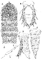 issued from : W. Lee & R. Huys in Zool. J. Linnean Soc., 2000, 129. [p.249, Fig.17]. Female: A, prosome, showing fine details of surface ornamentation (dorsal); B, urosome (ventral; caudal rami omitted); C; idem (lateral); E, right P6. Scale in microns. Nota: P5 very large, almost extending to postrior margin of anal somite, joining in ventral midline but not fused medially; uniramous, 1-segmented with vestigial suture line along inner margin marking boundary between protopod and exopod.
|
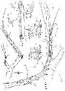 issued from : W. Lee & R. Huys in Zool. J. Linnean Soc., 2000, 129. [p.31, Fig.18]. Female: A1 (ventral); B, segment 7 of A1 (distal part; ventral); C, distal margin of A1 segment 3 (dorsal); D, anterior spinous process of A1 segment 2. male: E, A2 (anterior); F, free endopod of A2 (posterior; arrow indicating apical pore on short lateral seta); G, detail of distal armature of A2 (anterior). Nota: A1 7-segmented, with small sclerite around base of segment 1. A2 sexually dimorphic in allobasis, exopod and free endopod. Scales in microns.
|
 issued from : W. Lee & R. Huys in Zool. J. Linnean Soc., 2000, 129. [p.32, Fig.19]. Female: A, Md; B, right paragnath; C, Mx1 (posterior); D, Mx2 (anterior); E, Md (mandibular palp).. Scales in microns. Nota: Paragnaths well developed hirsute lobes. Mx1: praecoxa with transverse fold and few spinules around outer margin, arthrrite strongly developed with 2 large, swollen, plumose setae on anterior surface and 10 spines around distal margin; coxa with cylindrical endite bearing 2 bipinnate setae and 1 curved bipinnate spine; basis without discrete rami, apical margin not bilobate, elements grouped in inner cluster consisting of stout, curved pinnate spine and 2 bipinnate serae, and outer cluster consisting of 4 pinnate setae, increasing in length medially. Mx2 comprising syncoxa, allobasis and 3-segmented endopod
|
 issued from : W. Lee & R. Huys in Zool. J. Linnean Soc., 2000, 129. [p.33, Fig.20]. Female: A, A2; B, labrum (posterior); C, Mxp; D, distal armature of A2 endopod. Scales in microns. Nota: A2 3-segmented, comprising coxa, allobasis and free 1-segmented endopod; exopod 3-segmented; allobasis and endopod with numerous fine surface spinules. Labrum well developed, with elaborate spinular ornamentation along distal margin and numerous on posterior face. Mxp 2-segmented, comprising undivided protopod and 1-segmented endopod (without surface sutures marking original segmentation).
|
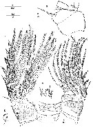 issued from : W. Lee & R. Huys in Zool. J. Linnean Soc., 2000, 129. [p.35, Fig.21]. Female: A, P1 endopod 2 (posterior; showing fusion of ancestral middle and distal segments); B, P1 (anterior); C, P4 (posterior). Male: D, P4 basis (medial lobate extension); E, P1 basis (anterior; surface ornamentation omitted). Scales in microns.
|
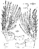 issued from : W. Lee & R. Huys in Zool. J. Linnean Soc., 2000, 129. [p.36, Fig.22]. Female: A, P2 (posterior); B, P3 (anterior). Male: CAD, medial lobe extension of bases P2-P3. Scales in microns.
|
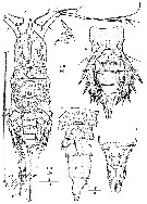 issued from : W. Lee & R. Huys in Zool. J. Linnean Soc., 2000, 129. [p.37, Fig.23]. Male: A, habitus (dorsal; arrow indication original position of seta I): B, rostral area (lateral); C, urosome (ventral; caudal rami omitted); D, idem (lateral); E, anal somite (dorsal); F, distal margin of caudal ramus (ventral). Scales in microns. Nota: Urosome 6-segmented, comprising pediger somite 5, genital somite and 4 abdominal somites P5 legs joining midventrally but not fused medially. P6 legs not fused medially, symmetrical. Each P6 with outernaked, middle pinnate and inner vestigial element; anterior surface with short spinules, inner distal margin with cluster of long setules. Anal somite medially constricted, dorsal anterior surface folded; anal opening probably not functional, anal operculum weakly developed, smooth, dorsal sensillae positioned anterior to anal opening.
|
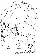
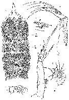 issued from : W. Lee & R. Huys in Zool. J. Linnean Soc., 2000, 129. [p.38, Fig.24]. Male: A, prosome showing fine details of surface ornamentation (dorsal); B, Md; C, mx1; D, Mx2 (posterior); E, Mx2 endopod (posterior; vestigial element arrowed); F, Mxp (tube pore arrowed). Scales in microns. Nota: Cephalosome produced into long spinous rostral projection, bearing several sensillae at its base. Labrum strongly folded. Paragnaths obsolete. Md completely atrophied, strongly reduced in size; gnathobase separated from rest of praecoxa by annulated constriction, with several pointed teeth around apical margin, 3 rudimentary structure of glandular nature discernible internally; palp minute, 1-segmented, tapering distally towards bifid apex, without setae. Mx1 completely atrophied, without distinct segment boundaries and strongly wrinkled; palp bent medially, with 4 vestigial setae; praecoxal arthrite with total of 12 setae; coxal endite drawn out into seta and bearing minute accessory seta. Mx2 consisting of syncoxa, allobasis and 3-segmented endopod; praecoxal endites absent; coxal endites minute lobes near boudary with allobasis, with 2 setal vestiges each; allobasis drawn out into slender, strongly curved, naked claw, accessory armature strongly reduced; endopod 3-segmented. Mxp 2-segmented, comprising protopod and endopod.
|
 issued from : W. Lee & R. Huys in Zool. J. Linnean Soc., 2000, 129. [p.40, Fig.25]. Male: A, A1 (ventral); B, P4 endopod; C, anterior spinous process on A1 segment 2; D, rudimentary segment 4 of A1 (dorsal). Scales in microns. Nota: A1 9-segmented, haplocer with geniculation between segments 7 and 8; segments 5 and 8 very elongate; segment 2 with small spinous process at anterior distal corner; segment 4 represented by small U-shaped sclerite.
| | | | | NZ: | 1 | | |
|
Distribution map of Scabrantenna yooi by geographical zones
|
| | | | | | | Loc: | | | Japan (Okinawa Trough, hydrothermal vent) | | | | N: | 1 | | | | Lg.: | | | (915) F: 3,53; M: 3,32; {F: 3,53; M: 3,32} | | | | Rem.: | epi-hyperbenthic (depths: 583-711 m).
For Lee & Huys (2000, p.41) this genus can be considered a transitionary genus between the more primitive genera, Nudivoras and Andromastax and the trudy planktonic Aegisthidae (Aegisthus mucronatusA. aculeatus. It shares with Aegisthus the strongly reduced mouthparts and the distally elongate A1 in the male. S. yooi is particularly reminiscent of A. aculeatus in the female morphology, such as the shape of the rostrum, the 7-segmented A1 and the form of Mxp. The most distinctive feature of the new genus is the A2 which displays a remarkable sexual dimorphism. A2 is clearly prehensile and capable of seizing; in view of the non feeding strategy of the male it is unlikely that A2 is involved in food manipulation and hence a role in mate guarding seems more likely. | | | Last update : 02/11/2008 | |
|
|
 Any use of this site for a publication will be mentioned with the following reference : Any use of this site for a publication will be mentioned with the following reference :
Razouls C., Desreumaux N., Kouwenberg J. and de Bovée F., 2005-2026. - Biodiversity of Marine Planktonic Copepods (morphology, geographical distribution and biological data). Sorbonne University, CNRS. Available at http://copepodes.obs-banyuls.fr/en [Accessed June 10, 2026] © copyright 2005-2026 Sorbonne University, CNRS
|
|
|
|


;)
;)
;)
;)
;)
;)
;)
;)
;)
;)

