|
|
 |
Fiche d'espèce de Copépode |
|
|
Harpacticoida ( Ordre ) |
|
|
|
Cervinioidea ( Superfamille ) |
|
|
|
Aegisthidae ( Famille ) |
|
|
|
Scabrantenna ( Genre ) |
|
|
| |
Scabrantenna yooi Lee & Huys, 2000 (F,M) | |
| | | | | | | Ref.: | | | Lee & Huys, 2000 (p.27, figs.F,M) | 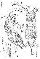 issued from : W. Lee & R. Huys in Zool. J. Linnean Soc., 2000, 129. [p.28, Fig.16]. Female (from Okinawa Trough): A, habitus (dorsal (distal 4/5 of caudal rami omitted); B, idem (lateral); C, caudal rami distal 4/5 (dorsal); D, ventral pore corresponding to original position of seta I (arrowed in A); E, seta II (dorsal); F, distal end of rami showing insertion sites of setae II-VII (dorsal).. Scales in microns. Nota: Prosome with complex surface reticulation consisting of anastomosing pattern of longutudinal and transversal lamellae; additional ornamentation consisting of sensillae and pores, paricularly around posterior margin of somites (Pediger 1 somite without sensillae, conspicuous aggregation of paired pores present middorsally near hind margin of cephalosome. 1st pediger somite separatd from dorsal cephalic shield, posterior margin smooth. Urosome 5-segmented. Anal somite with large anal opening, flanked by spinules laterally; anal operculum obsolete; dorsal sensillae positioned anterior to anal opening, ventral hind margin with large raised pores. Caudal rami slightly asymmetrical, covered with dense pattern of denticle-like spinules; each ramus with 6 setae, seta I absent, replaced by minute pore positioned asymmetrically on both rami in proximal quarter; seta II spiniform; seta III missing in all specimens examined but position indicated by large lateral scar (see the male); setae IV and V large; seta VI minute and displaced ventrally; seta VII presumably at base, positioned subterminally.
|
 issued from : W. Lee & R. Huys in Zool. J. Linnean Soc., 2000, 129. [p.249, Fig.17]. Female: A, prosome, showing fine details of surface ornamentation (dorsal); B, urosome (ventral; caudal rami omitted); C; idem (lateral); E, right P6. Scale in microns. Nota: P5 very large, almost extending to postrior margin of anal somite, joining in ventral midline but not fused medially; uniramous, 1-segmented with vestigial suture line along inner margin marking boundary between protopod and exopod.
|
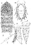
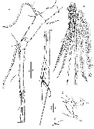
 issued from : W. Lee & R. Huys in Zool. J. Linnean Soc., 2000, 129. [p.31, Fig.18]. Female: A1 (ventral); B, segment 7 of A1 (distal part; ventral); C, distal margin of A1 segment 3 (dorsal); D, anterior spinous process of A1 segment 2. male: E, A2 (anterior); F, free endopod of A2 (posterior; arrow indicating apical pore on short lateral seta); G, detail of distal armature of A2 (anterior). Nota: A1 7-segmented, with small sclerite around base of segment 1. A2 sexually dimorphic in allobasis, exopod and free endopod. Scales in microns.
|
 issued from : W. Lee & R. Huys in Zool. J. Linnean Soc., 2000, 129. [p.32, Fig.19]. Female: A, Md; B, right paragnath; C, Mx1 (posterior); D, Mx2 (anterior); E, Md (mandibular palp).. Scales in microns. Nota: Paragnaths well developed hirsute lobes. Mx1: praecoxa with transverse fold and few spinules around outer margin, arthrrite strongly developed with 2 large, swollen, plumose setae on anterior surface and 10 spines around distal margin; coxa with cylindrical endite bearing 2 bipinnate setae and 1 curved bipinnate spine; basis without discrete rami, apical margin not bilobate, elements grouped in inner cluster consisting of stout, curved pinnate spine and 2 bipinnate serae, and outer cluster consisting of 4 pinnate setae, increasing in length medially. Mx2 comprising syncoxa, allobasis and 3-segmented endopod
|
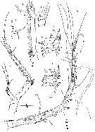
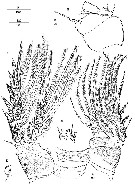
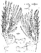
 issued from : W. Lee & R. Huys in Zool. J. Linnean Soc., 2000, 129. [p.33, Fig.20]. Female: A, A2; B, labrum (posterior); C, Mxp; D, distal armature of A2 endopod. Scales in microns. Nota: A2 3-segmented, comprising coxa, allobasis and free 1-segmented endopod; exopod 3-segmented; allobasis and endopod with numerous fine surface spinules. Labrum well developed, with elaborate spinular ornamentation along distal margin and numerous on posterior face. Mxp 2-segmented, comprising undivided protopod and 1-segmented endopod (without surface sutures marking original segmentation).
|
 issued from : W. Lee & R. Huys in Zool. J. Linnean Soc., 2000, 129. [p.35, Fig.21]. Female: A, P1 endopod 2 (posterior; showing fusion of ancestral middle and distal segments); B, P1 (anterior); C, P4 (posterior). Male: D, P4 basis (medial lobate extension); E, P1 basis (anterior; surface ornamentation omitted). Scales in microns.
|
 issued from : W. Lee & R. Huys in Zool. J. Linnean Soc., 2000, 129. [p.36, Fig.22]. Female: A, P2 (posterior); B, P3 (anterior). Male: CAD, medial lobe extension of bases P2-P3. Scales in microns.
|
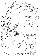
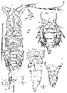 issued from : W. Lee & R. Huys in Zool. J. Linnean Soc., 2000, 129. [p.37, Fig.23]. Male: A, habitus (dorsal; arrow indication original position of seta I): B, rostral area (lateral); C, urosome (ventral; caudal rami omitted); D, idem (lateral); E, anal somite (dorsal); F, distal margin of caudal ramus (ventral). Scales in microns. Nota: Urosome 6-segmented, comprising pediger somite 5, genital somite and 4 abdominal somites P5 legs joining midventrally but not fused medially. P6 legs not fused medially, symmetrical. Each P6 with outernaked, middle pinnate and inner vestigial element; anterior surface with short spinules, inner distal margin with cluster of long setules. Anal somite medially constricted, dorsal anterior surface folded; anal opening probably not functional, anal operculum weakly developed, smooth, dorsal sensillae positioned anterior to anal opening.
|
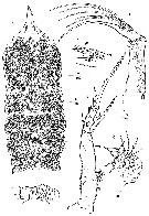 issued from : W. Lee & R. Huys in Zool. J. Linnean Soc., 2000, 129. [p.38, Fig.24]. Male: A, prosome showing fine details of surface ornamentation (dorsal); B, Md; C, mx1; D, Mx2 (posterior); E, Mx2 endopod (posterior; vestigial element arrowed); F, Mxp (tube pore arrowed). Scales in microns. Nota: Cephalosome produced into long spinous rostral projection, bearing several sensillae at its base. Labrum strongly folded. Paragnaths obsolete. Md completely atrophied, strongly reduced in size; gnathobase separated from rest of praecoxa by annulated constriction, with several pointed teeth around apical margin, 3 rudimentary structure of glandular nature discernible internally; palp minute, 1-segmented, tapering distally towards bifid apex, without setae. Mx1 completely atrophied, without distinct segment boundaries and strongly wrinkled; palp bent medially, with 4 vestigial setae; praecoxal arthrite with total of 12 setae; coxal endite drawn out into seta and bearing minute accessory seta. Mx2 consisting of syncoxa, allobasis and 3-segmented endopod; praecoxal endites absent; coxal endites minute lobes near boudary with allobasis, with 2 setal vestiges each; allobasis drawn out into slender, strongly curved, naked claw, accessory armature strongly reduced; endopod 3-segmented. Mxp 2-segmented, comprising protopod and endopod.
|
 issued from : W. Lee & R. Huys in Zool. J. Linnean Soc., 2000, 129. [p.40, Fig.25]. Male: A, A1 (ventral); B, P4 endopod; C, anterior spinous process on A1 segment 2; D, rudimentary segment 4 of A1 (dorsal). Scales in microns. Nota: A1 9-segmented, haplocer with geniculation between segments 7 and 8; segments 5 and 8 very elongate; segment 2 with small spinous process at anterior distal corner; segment 4 represented by small U-shaped sclerite.
| | | | | NZ: | 1 | | |
|
Carte de distribution de Scabrantenna yooi par zones géographiques
|
| | | | | | | Loc: | | | Japan (Okinawa Trough, hydrothermal vent) | | | | N: | 1 | | | | Lg.: | | | (915) F: 3,53; M: 3,32; {F: 3,53; M: 3,32} | | | | Rem.: | epi-hyperbenthic (depths: 583-711 m).
Voir aussi les remarques en anglais | | | Dernière mise à jour : 02/11/2008 | |
|
|
 Toute utilisation de ce site pour une publication sera mentionnée avec la référence suivante : Toute utilisation de ce site pour une publication sera mentionnée avec la référence suivante :
Razouls C., Desreumaux N., Kouwenberg J. et de Bovée F., 2005-2026. - Biodiversité des Copépodes planctoniques marins (morphologie, répartition géographique et données biologiques). Sorbonne Université, CNRS. Disponible sur http://copepodes.obs-banyuls.fr [Accédé le 18 juin 2026] © copyright 2005-2026 Sorbonne Université, CNRS
|
|
|
|


;)
;)
;)
;)
;)
;)
;)
;)
;)
;)

