|
|
 |
|
Calanoida ( Order ) |
|
|
|
Diaptomoidea ( Superfamily ) |
|
|
|
Acartiidae ( Family ) |
|
|
|
Acartia ( Genus ) |
|
|
|
Odontacartia ( Sub-Genus ) |
|
|
| |
Acartia (Odontacartia) edentata Srinui, Ohtsuka & Metillo, 2019 (F,M) | |
| | | | | | | Syn.: | Acartia (Odontacartia) pacifica sensu lato | | | | Ref.: | | | Srinui & al., 2019 (p.71, Descr. F, M, figs.M, fig.1, mol. biol, phylogeny, fig.7: range t° & Sal., key: p.89, 90); Lee S. & al., 2019 (p.86, Table 3). |  Remark: Erratum concerning the name: read Srinui and not Siruani. Female: 1 - Genital double-somite lacking posterodorsal sharp processes. 2 - Ventroposterior corners of prosome acutely pointed, reaching beyond half of ventral double-somite. Male: 1 - Urosomite 3 with large spine-like processes dorsally. 2 - Dorsal processes of urosomite 3 long, reaching half-length of anal somite. 3 - Urosomite 4 lacking spine-like processes between pair of dorsal processes. 4 - Genital somite with spinular rows along posterodorsal border. 5 - Inner projection of 1st exopodal segment of right P5 irregularly triangular.
|
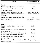 Issued from : K. Srinui, S. Ohtsuka, E.B. Metillo & M. Nishibori in ZooKeys, 2019, 814. [p.83,Table 2] Morphological characteristics in Acartia (Odontacartia) edentata. Compare with A. (O.) pacifica and A. (O.) ohtsukai, closely related species.
|
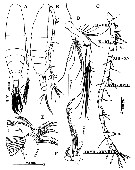 Issued from : K. Srinui, S. Ohtsuka, E.B. Metillo & M. Nishibori in ZooKeys, 2019, 814. [p.76, Fig.2]. Female (from 11°30'70''N, 124°69'01''E): A-B, habitus (dorsal and lateral, respectively); C, A1 (Roman numerals denote ancestral segment numbers); D, A2; E, Mx1; F, Mxp. Nota: - Cephalosome and 1st pediger separate, 4th and 5th pedigers fused. - Anterior margin of cephalosome triangular in dorsal view. - Rostrum with pair of thick, strong and sharp filaments. - Posterior prosome symmetrical with with pair of acute processes on each side: large ventrolateral, pointed processes with pair of small prrominences between and pair of smaller, pointed processes dorsally. - Urosome of 3 free somiyes. - Genital double-somite symmetrical with ratio of width-length approximately 1 : 1, lacking posterodorsal pointed processes. - 2nd urosomite with pair of strong, posterodorsal, pointed processes. - Anal somite as wide as long, without lateral rows of fine setules. - Proportional lengths of urosomites and caudal ramus 41 : 22 : 15 : 22 (= 100). - Caudal rami with setules along lateral margin, symmetrical, with 6 plumoses setae (II-VII). I absent, V longest and VII inserted anterodorsally. - A1 17-segmented, reaching beyond posterior end of 2nd urosomite, symmetical. Segments II-VIII completely fused; segments II, IV, VII, VIII, XI, XV and XVII with aesthetasc (ae). - A2: coxa with single seta; basis fused to elongated 1st endopodal segment forming allobasis with 8 setae on outer medial margin, and single lateral seta and trensverse row of small spinules terminally; 2nd segment with 8 outer setae and fine setules along inner margin; free terminal segment short with 6 setae. Exopod 3-segmented, satal formula: 1, 4, 3. - Mx1: praecoxal arthrite bearing 9 strong spines; coxal endite with 3 terminal setae; coxal epipodite with 1 short and 8 long setae; basal exite with 1 terminal seta and 1 proximal seta; exopod 1-segmented fused with basis and bearing 2 long medial setae, 5 setae terminally, with fine spinules along outer marginal; endopod absent. - Mxp: highly reduced; syncoxa with setation formula of 2 long, 1 medium and 1 short seta; basis with 1 short strong, 1 long setae and row of setules along inner margin; endopod 2-segmented, with 4 inner spines and terminal spiniform element.
|
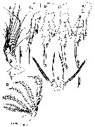 Issued from : K. Srinui, S. Ohtsuka, E.B. Metillo & M. Nishibori in ZooKeys, 2019, 814. [p.78, Fig.3]. Female: A, Md; B, Mx2; C, P1; D, P2; E, P3; F, P4; G, P5. - Md: gnathobase having 2 sharp cuspid teeth, 1 blunt tooth, and 3 small sharp teeth bordered by small spinules at the proximal end; basis with fine setules on medial outer marginal, single seta distally, and patch of small spinules on surface at midlength; 1st endopodal segment short with 2 short setae, 2nd segment with 11 setae; exopod 4-segmented, 1st to 4th with setal formula: 1, 1, 2, 2; 1st segment with row of small spinules. - Mx2: syncoxal endite bearing 5,3, 3, 2 setae; basis with 1 long seta; endopode with 4 long, 1 medium, and 1 short setae. - P1 to P4 biramous, each with 2-segmented endopod and 3-segmented exopod; coxa unarmed; 2nd endopodal segments of leg 1 and 4 and 3rd exopodal segment of leg 1 with row of small spinules anteriorly. - P5 symmetrical, coxae and intercoxal sclerite completely fused; basis longer than wide, outer margin with single lateral seta, slightly longer than terminal seta of exopod; exopod with knob-like projection basally, distal half spinulose.
|
 Issued from : K. Srinui, S. Ohtsuka, E.B. Metillo & M. Nishibori in ZooKeys, 2019, 814. [p.77, Table 1]. Female and Male: Armature formula for P1 to P4. Spines and setae indicated by Roman and Arabic numerals, respectively.
|
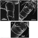 Issued from : K. Srinui, S. Ohtsuka, E.B. Metillo & M. Nishibori in ZooKeys, 2019, 814. [p.81, Fig.5]. SEM micrographs of female. A, Genital double-somite (lateral view); B, urosomite (ventral view); C, Genital double-somite ( ventral view). Nota: Paired genital slits are located at midlength and moderately separated (fig. B, C. A row of setules is located along the anterior margin of each genital slit (fig.C)
|
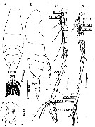 Issued from : K. Srinui, S. Ohtsuka, E.B. Metillo & M. Nishibori in ZooKeys, 2019, 814. [p.80, Fig.4]. Male: A-B, habitus (dorsal and lateral, respectively); C, left A1; D, right A1. (Roman numerals denote ancestral segment numbers). E, rostrum of male; F, rostrum of female. Nota: - Body similar to that of female. - Cephalosome anterior bluntly triangular in dorsal view. - Rostrum with paired filaments, slightly curved (versus straight in female). - Posterior prosome symmetrical with pair of short acute processes dorsolaterally and longer ventrolateral acute processes, with pair of small prominences between 2 dorsolateral processes. Postrior margin of prosome naked. - Urosome of 5 somites, symmetrical in dorsal view. - Genital somite (1st urosomite) as long as wide, bearing 2 dorsolateral rows of small spinules. - 2nd urosomite with 2 pairs of strong posterior dorsolateral processes, outer shorter than inner, and furnished with 3 rows of minute spinules ventrolaterally. - 3rd urosomite with pair of strong acute processes dorsally; . - 4th urosomite 4.5 times shortert han wide and furnished with pair of small medium-sized acute processes dorsally. - Anal somite with setules along outer margins. - Caudal rami symmetrical, approximately 1.5 times as long as wide, having lateral setules along inner margin , 6 plumose (II-VII) setae as in female. - Left A1 incompletely 21-segmented; segments 2, 3 and 21 incompletely fused. Right A1 geniculate, incompletely 17-segmented, not reaching beyond posteriorv end of 5th pedigerous somite; segments 2 and 3 incompletely fused; segments 2, 3, 4, 7, 9, 15, 17 each with aesthetasc (ae); segments 4, 7 and 12 each with spiniform element.
|
 Issued from : K. Srinui, S. Ohtsuka, E.B. Metillo & M. Nishibori in ZooKeys, 2019, 814. [p.78, Fig. 3, H]. Male: H, P5. Nota: P5 uniramous; coxae unarmed and completely fused with intercoxal sclerite; each side of basis with outer plumose seta subterminally, left basis about 2.5 times as lo,g as wide, with concave inner margin. Right exopod 3-segmented, 1st segment about 3 times as long as wide with single seta subterminally, 2nd segment with small subterminal spine at mid-length of inner irregularly triangular knobs, 3rd segment curved inward with small terminal spine and small inner spine midway. left exopod 2-segmented, 1st segment about 2.5 times as long as wide, 2nd segment with long inner seta and small terminal spine.
|
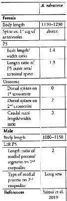 Issued from : S. Lee, H.Y. Soh & W. Lee in ZooKeys, 2019, 893. [p.84, Table 3]. Acartia (Odontacartia) edentata: Morphological characters. Compare with other Odontacartia species. Nota: 1 - Absence of spine on 1st to 2 nd segments of female A1 .....2. 2 - Dorsal surface of female genital double-somite devoid of processes (spines and spinules) ....... 3. 3 - Female caudal rami three times longer than wide; medial process and 2nd exopodite segment of male left P5 twice longer than 2nd exopodite segment.
| | | | | NZ: | 1 | | |
|
Distribution map of Acartia (Odontacartia) edentata by geographical zones
|
| | | 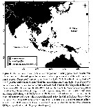 Issued from : K. Srinui, S. Ohtsuka, E.B. Metillo & M. Nishibori in ZooKeys, 2019, 814. [p.73, Fig. I]. Issued from : K. Srinui, S. Ohtsuka, E.B. Metillo & M. Nishibori in ZooKeys, 2019, 814. [p.73, Fig. I]. |
| | | | Loc: | | | Philippines (Carigara Bay, off Leyte Island)
11°30'70'' N, 124°69'01'' E. | | | | N: | 1 | | | | Lg.: | | | (1224)F: 1,19-1,23; M: 1,08-1,15; {F: 1,19-1,23; M: 1,08-1,15} | | | | Rem.: | The species belongs to sub-group centrura from Acartia (Odontacartia).
Among the centrura species, the female of A/ (O.) edentata is unique in lacking paired posterior dorsolateral processes on the genital double-somite unlike those of the closely related A. (O.) pacifica from Japan and Korea, and O. ohtsukai according to Ueda & Bucklin (2006).
For Srinui & al. (2019, p.84) in the tropical zone, A. (O.) edentata collected in the Philippines during the rainy season (August), when water temperature and salinity were 30.2 °C and 33.2 per 1000, respectively, in contrast A. (Acanthacartia) tsuensis represents the dominant species in brackish pond water from Panay Island §central Philippines) during the dry season (November-April), with salinity ranging from 14.0 to 40.0. The authors conclude that three species of Acartia (A. (O.) ohtsukai, A/ (O.) pacifica, A. (O.) edentata, and probably other Acartia species appear to occupy water bodies differing in temperature and salinity of the tropical and subtropical zone of the Pacific and Indian oceans. | | | Last update : 09/03/2020 | |
|
|
 Any use of this site for a publication will be mentioned with the following reference : Any use of this site for a publication will be mentioned with the following reference :
Razouls C., Desreumaux N., Kouwenberg J. and de Bovée F., 2005-2025. - Biodiversity of Marine Planktonic Copepods (morphology, geographical distribution and biological data). Sorbonne University, CNRS. Available at http://copepodes.obs-banyuls.fr/en [Accessed December 28, 2025] © copyright 2005-2025 Sorbonne University, CNRS
|
|
 |
 |


