|
|
 |
|
Calanoida ( Order ) |
|
|
|
Clausocalanoidea ( Superfamily ) |
|
|
|
Clausocalanidae ( Family ) |
|
|
|
Clausocalanus ( Genus ) |
|
|
| |
Clausocalanus lividus Frost & Fleminger, 1968 (F,M) | |
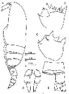
| | | | | | | Syn.: | Clausocalanus arcuicornis sp.2 : Fleminger,1964 a; 1967 a (tab. I) | | | | Ref.: | | | Frost & Fleminger, 1968 (p.35, figs.F,M, Rem.); Carli & Crisafi, 1969 (p.277, figs.F,M); Razouls, 1972 (p.94, Annexe: p.31, 33, figs.F, Rem.); Williams & Wallace, 1975 (p.175, fig.F); Dawson & Knatz, 1980 (p.7, 8, figs.F,M); Alvarez-Marques, 1981 (p.157, figs.F, Rem.); Gardner & Szabo, 1982 (p.182, figs.F,M); Bradford-Grieve, 1994 (p.118, figs.F,M, fig.101); Chihara & Murano, 1997 (p.776, Pl.90,93: F,M); Bradford-Grieve & al., 1999 (p.878, 916, figs.F,M); Bucklin & al., 2003 (p.335, tab.2, fig.1, Biomol); Blanco-Bercial & Alvarez-marqués, 2007 (p.73, fig.2, Biomol.); Avancini & al., 2006 (p.78, Pl. 47, figs.F,M, Rem.); Vives & Shmeleva, 2007 (p.619, figs.F,M, Rem.); Blanco-Bercial & al., 2011 (p.108, fig.3, phylogeographic biomol. analysis); Blanco-Bercial & al., 2014 (p.5: Rem.: problematic taxa); | 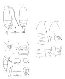 issued from : B. Frost & A. Fleminger in Bull; Scripps Inst. Oceanogr. Univ. California, 1968, 12. [Pl.14, p.128-129; Pl.15, p.130-131; Pl.16, p.132-133]. Female: 1 a, habitus (right lateral view); 1 b, idem (dorsal view); taken from different specimens.2 a, b, frontal region (right lateral view); 2, c-f, rostrum (right lateral view); 2 g, rostrum (anterior view); 2, h-j, Th. 4-5 (posterior part) and genital segment (right lateral view); from different specimens; 3 a, Th.4-5 (posterior part) and genital segment (right lateral view); 3 b, genital segment (ventral view); 3 c, Th.4-5 (posterior part) and urosome with spermatophore attached (right lateral view); 3 d, B2 of P2; 3 e, B2 of P3; 3 f, P5; from different specimens.
|
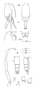 issued from : B. Frost & A. Fleminger in Bull; Scripps Inst. Oceanogr. Univ. California, 1968, 12. [Pl.17, p.134-135; Pl.18, p.136-137]. Male: 1 a, habitus (right lateral view); 1 b, idem (dorsal view); 1 c, frontal region (right lateral view); 1 d, urosome 4 (posterior part), urosome 5 and caudal rami (armature incomplete) (dorsal view); from different specimens. 2 a, Th.4-5 (posterior part) and urosome (right lateral view); 2 b, urosome (armature of caudal rami incomplete) (dorsal view); 2 c, B2 of P2; 2 d, B2 of P3; 2 e, P5 (right lateral view); 2 f, right P5 (posterior ); 2 g, right P5 (left lateral); from different specimens. Nota: 2nd segment of P5 more than 4.4 times as long as wide. Urosomal segment 2 more than 1.35 times as long as 2nd segment of P5.
|
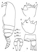
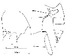
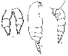
 issued from : J.M. Bradford-Grieve in The Marine Fauna of New Zealand: Pelagic Calanoid Copepoda. National Institute of Water and Atmospheric Research (NIWA). New Zealand Oceanographic Institute Memoir, 102, 1994. [p.118, Fig.67]. Female: A, habitus (lateral right side); B, genital somite (lateral right side); C, rostrum (lateral view); D, basipod of P3. Male: F, habitus (lateral left side); G, basipod 2 of P3; H, P5.
|
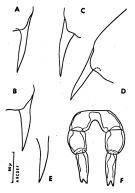
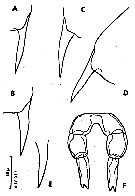
 issued from : C. Razouls in Thèse Doc. ès Sciences, Univ. P & M. Curie, Paris , Tome annexe, 1972, Fig.32 (unpublished). Female (from Banyuls): A, habitus (lateral left side); B, basipod 2 of P2; C, basipod 2 of P3; D, P5; E, anal segment and right furcal ramus (dorsal).
|
 issued from : C. Razouls in Thèse Doc. ès Sciences, Univ. P & M. Curie, Paris , Tome annexe, 1972, Fig.33 (unpublished). Female (from Banyuls): A-E, rostrum from different specimens (lateral view); F, P5.
|
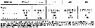 issued from : B. Frost & A. Fleminger in Bull. Scripps Inst. Oceanogr. Univ. California, 1968, 12. [p.30, Table 2a]. Clausocalanus lividus females: Measurements and ratios *.
|
 issued from : B. Frost & A. Fleminger in Bull. Scripps Inst. Oceanogr. Univ. California, 1968, 12. [p.31, Table 2b]. Clausocalanus lividus males: Measurements and ratios *. r = sample range; m = sample mean; n = number of specimens measured; s = sample standard deviation.
|
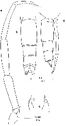
 issued from : B. Frost & A. Fleminger in Bull. Scripps Inst. Oceanogr. Univ. California, 1968, 12. [p.102, P1.1, b, c]. Female: b, urosome with spermatophore attached (right lateral); c, basipodite 2 of P3. cm = cementing material; am = accessory material associated with spermatophore neck; sn = spermatophore neck; ss = spermatophore sac; D = proximal width of basipodite 2 spiniform process 3; L = distance between processes 2 and 3.
|
 issued from : B. Frost & A. Fleminger in Bull. Scripps Inst. Oceanogr. Univ. California, 1968, 12. [p.27, Fig.10]. Urosomal segment 2:segment 2 of P5 length ratio (ordinate) plotted against urosomal segment 2 length tabscissa) for males of Clausocalanus lividus and C. mastigophorus.
|
 issued from : B. Frost & A. Fleminger in Bull. Scripps Inst. Oceanogr. Univ. California, 1968, 12. [p.18, Fig.1]. Female caudal ramus length-to-width ratio (ordinate) plotted against caudal ramus length (abscissa) for Clausocalanus mastigophorus and C. lividus.
|
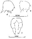
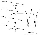 issued from : B. Frost & A. Fleminger in Bull. Scripps Inst. Oceanogr. Univ. California, 1968, 12. [p.131, Pl.15, c-f, g]. Female: c-f, rostrum (right lateral); g, rostrum (anterior).
|
 issued from : B. Frost & A. Fleminger in Bull. Scripps Inst. Oceanogr. Univ. California, 1968, 12. [p.133, Pl.16, d, e, f]. Female: d, basipodite 2 of P2; e, basipodite 2 of P3; f, P5.
|
 issued from : B. Frost & A. Fleminger in Bull. Scripps Inst. Oceanogr. Univ. California, 1968, 12. [p.137, Pl.18, a, b, e, f-g]. Male: a, posterior part of last thoracic segment and urosome (right lateral); b, urosome (dorsal); e, P5 (right lateral); f-g, right P5 (left lateral).
|
 issued from : B. Frost & A. Fleminger in Bull. Scripps Inst. Oceanogr. Univ. California, 1968, 12. [p.137, Pl.18, c, d]. Male: c, basipodite 2 of P2; d, basipodite 2 of P3.
|
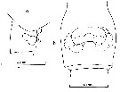 issued from : B. Frost & A. Fleminger in Bull. Scripps Inst. Oceanogr. Univ. California, 1968, 12. [p.133, Pl.16, a, b]. Female: a, posterior part of last thoracic segment and genital segment (right lateral); b, genital segment (ventral; see legend of structure of seminal receptacle in C. arcuicornis).
|
 issued from : C. Razouls in Th. Doc. Etat Fac. Sc. Paris VI, 1972, Annexe. [Fig.32]. Female (from Banyuls, G. of Lion): A, habitus (lateral); B, basipod 2 of P2; C, basipod 2 of P3; D, P5; E, anal segment and left caudal ramus (dorsal).
|
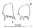
 issued from : C. Razouls in Th. Doc. Etat Fac. Sc. Paris VI, 1972, Annexe. [Fig.34]. Female: A-E, various forms of rostrum; F, P5.
|
 issued from : C. Razouls in Th. Doc. Etat Fac. Sc. Paris VI, 1972, Annexe. [Table XXXI] (unpublished). Sizes and ratios in Female (from Banyuls, G. of Lion): Lt= Bodyl length; C= Cephalolome length; P= prosome length; U= urosome length; U1= 1st urosomal segment; U2= 2nd urosomal length; U3= 3rd urosomal segment; F= caudal ramus length; n= number of specimens; moyenne= mean; Cv= coefficient of variation (percentage).
|
 Issued from F. Alvarez-Marques in Rev. Fac. Cienc. Univ. Oviedo (Ser. Biologia), (1979-80), 20-21, 1981. [p.161, Pl. I, Figs.3-5]. Female (from Gijon, NW Spain): 3, habitus (lateral); 4, urosome and spermatheca (lateral); 2, P5.
| | | | | Compl. Ref.: | | | Björnberg, 1973 (p.310, 385); Carter, 1977 (1978) (p.35); Kovalev & Schmeleva, 1982 (p.83); Scotto di Carlo & Ianora, 1983 (p.150); Scotto di Carlo & al., 1984 (p.1041); Tremblay & Anderson, 1984 (p.6: Rem.); Regner, 1985 (p.11, Rem.: p.27); Brenning, 1985 a (p.28, Table 2); Greze & al., 1985 (p.7); Hure & Krsinic, 1998 (p.100); Pancucci-Papadopoulou & al., 1990 (p.199); Ianora & al., 1990 (p.249, fig., parsitism effects); Scotto di Carlo & al., 1991 (p.270); Seguin & al., 1993 (p.23); Saiz & Calbet, 1999 (p.599, reproduction); Moraitou-Apostolopoulou & al., 2000 (tab.I, fig.6); Fragopoulu & al., 2001 (p.49, tab.1); Mackas & Galbraith, 2002 a (p.423, Table 2); Vukanic, 2003 (139, tab.1); Hsiao & al., 2004 (p.325, tab.1); Lan & al., 2004 (p.332, tab.1); Peralba & Mazzocchi, 2004 (p.645, figs.3,6); Lo & al., 2004 (p.89, tab.1); Vukanic & Vukanic, 2004 (p.9, tab. 2, 3); Fernandez & al., 2004 (p.501, tab.5); Isari & al., 2006 (p.241, tab.II); Hooff & Peterson 2006 (p.2610); Valdés & al., 2007 (p.103: tab.1); Dur & al., 2007 (p.197, Table IV); Khelifi-Touhami & al., 2007 (p.327, Table 1); Cabal & al., 2008 (289, Table 1); Lan Y.C. & al., 2008 (p.61, Table 1, % vs stations, Table 2: indicator species, Rem.: p.72); Galbraith, 2009 (pers. comm.); Lan Y.-C. & al., 2009 (p.1, Table 2, % vs hydrogaphic conditions); Brugnano & al., 2010 (fig.8); Schnack-Schiel & al., 2010 (p.2064, Table 2: E Atlantic subtropical/tropical, Fig.4, 6, 7, Tabe 4); Mazzocchi & Di Capua, 2010 (p.425); Hsiao S.H. & al., 2011 (p.475, Appendix I); Hsiao & al., 2011 (p.317, Table 2, indicator of seasonal change); Isari & Saiz, 2011, feeding); Mazzocchi & al., 2011 (p.1163, Table II, long-term time-series 1984-2006); Mazzocchi & al., 2012 (p.135, annual abundance 1984-2006); Uysal & Shmeleva, 2012 (p.909, Table I); Miloslavic & al., 2012 (p.165, Table 2, transect distribution); Takahashi M. & al., 2012 (p.393, Table 2, water type index); Brugnano & al., 2012 (p.207, Table 3); in CalCOFI regional list (MDO, Nov. 2013; M. Ohman, comm. pers.); Hirai & al., 2013 (p.1, Table I, molecular marker); Peijnenburg & Goetze, 2013 (p.2765, genetic data); Lidvanov & al., 2013 (p.290, Table 2, % composition); Fernandez de Puelles & al., 2014 (p.82, Table 3, seasonal abundance); Mazzocchi & al., 2014 (p.64, Table 5, abundance); Benedetti & al., 2016 (p.159, Table I, fig.1, functional characters); Benedetti & al., 2018 (p.1, Fig.2: ecological functional group) ; Belmonte, 2018 (p.273, Table I: Italian zones); Hirai & al., 2020 (p.1, Fig. 5: cluster analysis (OTU), spatial distribution). | | | | NZ: | 15 | | |
|
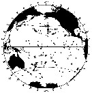
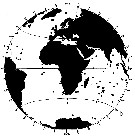
Distribution map of Clausocalanus lividus by geographical zones
|
| | | | | | | | | | | |  issued from : B. Frost & A. Fleminger in Bull. Scripps Inst. Oceanogr. Univ. California, San Diego, 1968, 12. [p.36, Chart 2, a]. issued from : B. Frost & A. Fleminger in Bull. Scripps Inst. Oceanogr. Univ. California, San Diego, 1968, 12. [p.36, Chart 2, a].
Occurrence of C. lividus in samples examined; closed circles represent samples examined; open circles represent samples in which adults were found; bars through open circles represent samples from which specimens were removed for measurements. |
 issued from : B. Frost & A. Fleminger in Bull. Scripps Inst. Oceanogr. Univ. California, San Diego, 1968, 12. [p.37, Chart 2, b]. issued from : B. Frost & A. Fleminger in Bull. Scripps Inst. Oceanogr. Univ. California, San Diego, 1968, 12. [p.37, Chart 2, b].
Occurrence of C. lividus in samples examined; closed circles represent samples examined; open circles represent samples in which adults were found; bars through open circles represent samples from which specimens were removed for measurements. |
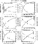 Issued from : S. Isari & E. Saiz in J. Plankton Res., 2011, 33 (5). [p.719, Fig.1]. Issued from : S. Isari & E. Saiz in J. Plankton Res., 2011, 33 (5). [p.719, Fig.1].
Functional feeding of Clausocalanus lividus female on six different-sized food items. Clearance rates (ml./copepode/day) and injestion rates (cells/copepode/day) are presented as a function of the average food concentration (cells/ml.). Error bars indicate the standard errors of the mean values.
Nota: Live copepods were collected at approximately five nautical miles from the coast of Barcelona (Spain), ca. 100 m depth, from April to June 2009. |
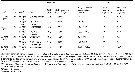 Issued from : S. Isari & E. Saiz in J. Plankton Res., 2011, 33 (5). [p.717, Table 1]. Issued from : S. Isari & E. Saiz in J. Plankton Res., 2011, 33 (5). [p.717, Table 1].
Overview of the experiments concerning the food of Clausocalanus lividus. |
| | | | Loc: | | | China Seas (East China Sea, South China Sea), Taiwan Strait, Taiwan (SW, E, NW, N: Mienhua Canyon), Japan, off SE Japan, Pacif. (N & S subtropical & temperate), Gulf of Alaska (58°N, 147°W), Vancouver Is., off Washington coast, Oregon (off Newport), California, New Zealand, Tahiti, Chile, Indian S, E South Africa (Natal), off S Cape Verde Is., Morocco-Mauritania, G. of Mexico, Florida, New-York, Gulf Stream (41° N,58° W), off S Newfoundland, off W Ireland, Bay of Biscay, Gijon (NW Spain), Cantabrian Sea, Ibero-moroccan Bay, Medit. (Alboran Sea, Gulf of Annaba, Baleares, off Barcelona coast, Banyuls, Ligurian Sea, Tyrrhenian Sea, G. of Naples, Strait of Messina, Adriatic Sea, Ionian Sea, Aegean Sea, Lebanon Basin) | | | | N: | 70 | | | | Lg.: | | | (30) F: 1,77-1,26; M: 1,45-1,13; (374) F: 1,75-1,262; (784) F: 1,98-1,66; (1138): F: 1,79-1,22; M: 1,30-1,09; {F: 1,22-1,98; M: 1,09-1,45}
The mean female size is 1.587 mm (n = 8; SD = 0.2948) and the mean male size is 1.243 mm (n = 4; SD = 0.1658). The size ratio (male : female) is about 0.82 | | | | Rem.: | epipelagic. For Frost & Fleminger (1968, p.38) C. lividus female is distinguished from the very similar ingens by the shape and curvature of the forehead and rostrum in lateral view. C. lividus and laticeps differ in the inclination of the rostrum in lateral view, the length-width ratio of the caudal ramus, the form and spacing of basipodite 2 spiniform processes 2 and 3 of P3, and usually in the shape of the frontal region of the anterior portion of body including the cephalosome and fusedthoracic segment bearing P1, in lateral and dorsal view. C. lividus female differs from jobei and minor in the form of the rostrum in lateral view. The male of C. lividus is distinguished from ingens by the Prosome/Urosomal segment 2 length ratio and the length-width ratio of the left 2nd segment of P5. C. lividus and males differ in the urosomal segment 2/2nd segment of P5 and urosomal segment 3/3rd segment of P5 length ratios, in the length-width ratio of the caudal ramus, and in the form of basipodite 2 spiniform processes 2 and 3 of P3. The name lividus refers to the blue or lavender tint observed in the cuticle of late copepodid and adult specimens.
See in the annex (in Razouls, 1972, p.30-32) the comparison with the females of this species and Clausocalnus mastigophorus, notably the variability of the rostrum (figs31 and 33).
After Benedetti & al. (2018, p.1, Fig.2) this species belonging to the functional group 4 corresponding to small filter feeding herbivorous and mixed feeding omnivorous (mostly broadcasters). | | | Last update : 04/12/2020 | |
|
|
 Any use of this site for a publication will be mentioned with the following reference : Any use of this site for a publication will be mentioned with the following reference :
Razouls C., Desreumaux N., Kouwenberg J. and de Bovée F., 2005-2026. - Biodiversity of Marine Planktonic Copepods (morphology, geographical distribution and biological data). Sorbonne University, CNRS. Available at http://copepodes.obs-banyuls.fr/en [Accessed June 24, 2026] © copyright 2005-2026 Sorbonne University, CNRS
|
|
|
|


;)
;)
;)
;)
;)
;)
;)
;)
;)
;)
;)
;)
;)
;)
;)
;)
;)

;)
;)
;)
;)
