|
|
 |
Fiche d'espèce de Copépode |
|
|
Calanoida ( Ordre ) |
|
|
|
Diaptomoidea ( Superfamille ) |
|
|
|
Pontellidae ( Famille ) |
|
|
|
Labidocera ( Genre ) |
|
|
| |
Labidocera farrani Greenwood & Othman, 1979 (F,M) | |
| | | | | | | Syn.: | Labidocera sp. (M) Farran, 1936 a (p.116, figs.M);
L. detruncata : Dakin & Colefax, 1933 (p.206);
L. detruncatum : Dakin & Colefax, 1940 (var. de Sydney) (p.103, part. figs.F,M);
Labidocera sp. Greenwood, 1979 (p.103) | | | | Ref.: | | | Greenwood & Othman, 1979 (p.231, 237, figs.F,M); Kimmerer & al., 1985 (p.428); Bradford-Grieve, 1999 b (p.192, figs.F,M, Rem., figs.184, 194); Mulyadi, 2014 (p.1629, Table II) | 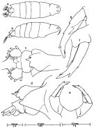 issued from : J.G. Greenwood in Proc. R. Soc. Qd, 1979, 90. [p.104, Fig.8]. As Labidocera sp. Female (from Moreton Bay, E Australia): a, habitus (dorsal); b, idem (lateral right side); c, urosome (dorsal); d, posterior metasome and urosome (lateral right side); e, P5; f, g, detail of left and right P5.
|
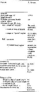
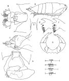 issued from : J.G. Greenwood & B.H.R. Othman in J. Plankton Res., 1979, 1 (3). [p.233, Fig.2]. Female (G. of Carpentaria): A, habitus (lateral right side); B, posterior metasomal border and urosome (dorsal); C, idem (lateral right side); D, rostrum; E, F, Md (medial and lateral surfaces of masticatory edge); G, P5 (posterior; H, urosome with outline of attached spermatophore and coupler (dorsal). Nota: The length ratio of the prosome to the urosome is 4.38:1. The dorsal eye-lenses are relatively small and are separated by more than one eye diameter. Urosome small, 3-segmented. Spermatophores were very common; the body of the sperm chamber is mounted dorsally on the urosome posterior to the genital segment.
|
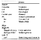
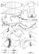 issued from : J.G. Greenwood & B.H.R. Othman in J. Plankton Res., 1979, 1 (3). [p.232, Fig.1]. Male (from G. of Carpentaria): A, habitus (dorsal); B, idem (lateral right side); C, posterior metasomal border and urosome (dorsal); D, rostrum; E, right A1; F, G, opposite views of right A1 segments 17-21; H, I, Md (medial and lateral surfaces of masticatory edge); J, left P5 (anterior); K, left P5 (detail of terminal segment); L, right P5 (anterior); M, right P5 (chela, semi lateral view showing detail of \"thumb\" and \"finger\"). Nota: The length ratio of the prosome to the urosome is 3.1:1. Head without lateral hooks but with lateral prominence at the bases of the A1. The two dorsal eye-lenses are large and almost contiguous. Urosome 5-segmented (considerable variation was found in the extent to which segments telescoped within those preceding.
|
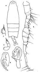 Issued from : G.P. Farran in Great Barrier Reef Expedition 1928-1929, Scient. Rep., V, N°3, 1936. [p.117, Fig.20]. As Labidocera sp. Male: a, habitus (dorsal); b, A1; c, right P5; d, the same (another view); e, left P5; f, terminal segment of same.
|
 issued from : J.G. Greenwood & B.H.R. Othman in J. Plankton Res., 1979, 1 (1). [p.237] Characters in L. farrani compared with other species within the super-species detruncata (see L. cervi, L. tasmanica, L. caudata, L. detruncata, L. pavo, L. bataviae, L. madurae and L. sinolobata, with reference sources).
|
 Issued from : Mulyadi in Crustaceana, 2014, 87 (14). [p.1630, Table II]. Characteristic features of L. farrani compared with five species in the sub-groups in Indo-Australian region. Compare with L. cervi, sinilobata, kaimanaensis and detruncata
| | | | | Ref. compl.: | | | Othman & al., 1990 (p.561, 564, Table 1) | | | | NZ: | 3 | | |
|
Carte de distribution de Labidocera farrani par zones géographiques
|
| | | | | | | Loc: | | | Australia (Shark Bay, Sydney, Moreton Bay, Great Barrier Reef, G. of Carpentaria) | | | | N: | 7 | | | | Lg.: | | | (34) M: 3,18-3,12; (104) F: 3,5; M: 2,8; (106) F: 3,22-2,82; M: 2,96-2,59; {F: 2,82-3,50; M: 2,59-2,96} | | | | Rem.: | épipélagique, néritique.
Voir aussi les remarques en anglais | | | Dernière mise à jour : 16/03/2018 | |
|
|
 Toute utilisation de ce site pour une publication sera mentionnée avec la référence suivante : Toute utilisation de ce site pour une publication sera mentionnée avec la référence suivante :
Razouls C., Desreumaux N., Kouwenberg J. et de Bovée F., 2005-2026. - Biodiversité des Copépodes planctoniques marins (morphologie, répartition géographique et données biologiques). Sorbonne Université, CNRS. Disponible sur http://copepodes.obs-banyuls.fr [Accédé le 07 mai 2026] © copyright 2005-2026 Sorbonne Université, CNRS
|
|
|
|


;)
;)
;)
;)
;)
;)

