|
|
 |
Fiche d'espèce de Copépode |
|
|
Calanoida ( Ordre ) |
|
|
|
Diaptomoidea ( Superfamille ) |
|
|
|
Pontellidae ( Famille ) |
|
|
|
Pontellopsis ( Genre ) |
|
|
| |
Pontellopsis villosa Brady, 1883 (F,M) | |
| | | | | | | Syn.: | Monops villosus : Giesbrecht, 1892 (p.486, 497, 773, figs.F,M); 1894;
Monops edwardsii (part.) Claus, 1893 | | | | Ref.: | | | Brady, 1883 (p.86, Descr.F, figs.F); Thompson, 1888 d (p.143); Giesbrecht & Schmeil, 1898 (p.148, Rem. F,M); A. Scott, 1909 (p.172, Rem.); Sars, 1925 (p.355); Sewell, 1932 (p.390); Rose, 1933 a (p.264, figs.F,M); Wilson, 1942 a (p.204, fig.M); C.B. Wilson, 1950 (p.314, fig.M); Voronina, 1962 a (p.68, 69, 73, fig.1, 5, 6); Tanaka, 1964 c (p.268, figs.M, No figs. F); Chen & Zhang, 1965 (p.108, figs.F,M); Vervoort, 1965 (p.194, Rem.); Owre & Foyo, 1967 (p.99, figs.F,M); Corral Estrada, 1970 (p.205, Rem., fig.F); Silas & Pillai, 1973 (1976) (p.837, figs.F,M, Rem.); Pillai, 1975 (p.137, figs.F,M, Rem.); 1977 (1982) (p.66); Björnberg & al., 1981 (p.660, figs.F,M); Zheng & al., 1982 [p.87, Figs.F,M); Ohtsuka & Onbé, 1991 (p.214); Ianora & al., 1992 (p.401, fig.); Chihara & Murano, 1997 (p.872, Pl.157,159: F,M); Bradford-Grieve & al., 1999 (p.885, 960, figs.F,M); Mulyadi, 2002 (p.151, figs.F, Rem.); Vives & Shmeleva, 2007 (p.519, figs.F,M, Rem.); Suarez-Morales & Kozak, 2012 (p.1, key F,M: p.14, fig.F,M). | 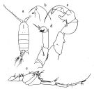 issued from : O. Tanaka in Publs Seto Mar. Biol. Lab., 1964, XII (3). [p.268, Fig.240]. Male: a, habitus (dorsal); b, forehead (left lateral side); c, clasping of right A1; d, left P5; e, right P5.
|
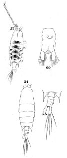 Issued from : W. Giesbrecht in Systematik und Faunistik der Pelagischen Copepoden des Golfes von Neapel und der angrenzenden Meeres-Abschnitte. – Fauna Flora Golf. Neapel, 1892. Atlas von 54 Tafeln. [Taf.41, Figs.45, 51, 57, 69]. As Monops villosus. Female: 57, habitus (dorsal); 69, urosome (ventral). Male: 45, last thoracic segment and urosome (dorsal); 51, habitus (dorsal).
|
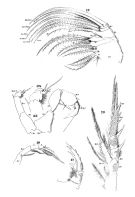 iIssued from : W. Giesbrecht in Systematik und Faunistik der Pelagischen Copepoden des Golfes von Neapel und der angrenzenden Meeres-Abschnitte. – Fauna Flora Golf. Neapel, 1892. Atlas von 54 Tafeln. [Taf.26, Figs. 10, 12, 17, 23, 33, 34]. As Monops villosus. Female: 17, P5 (posterior view). Male: 10, Mxp (extremity, posterior view); 12, Mx2 (posterior view); 23, P3 (posterior view); 33, P5; 34, P5 (distal part of left leg).
|
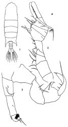 issued from: Q.-c Chen & S.-z. Zhang in Studia Marina Sinica, 1965, 7. [Pl.48, 1-3]. Male (from E China Sea): 1, habitus (dorsal); 2, right A1; 3, P5 (posterior).
|
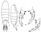 issued from: Q.-c Chen & S.-z. Zhang in Studia Marina Sinica, 1965, 7. [Pl.47, 8-11]. Female: 8, habitus (dorsal); 9, idem (lateral right side); 10, P5 (posterior); 11, right P5 (another specimen), posterior.
|
 issued from: Q.-c Chen & S.-z. Zhang in Studia Marina Sinica, 1965, 7. [Pl.47, 8-11]. Female: 8, habitus (dorsal); 9, idem (lateral right side); 10, P5 (posterior); 11, right P5 (another specimen), posterior.
|
 issued from: Q.-c Chen & S.-z. Zhang in Studia Marina Sinica, 1965, 7. [Pl.47, 8-11]. Female: 8, habitus (dorsal); 9, idem (lateral right side); 10, P5 (posterior); 11, right P5 (another specimen), posterior.
|
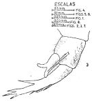 issued from : J. Corral Estrada in Tesis Doct., Univ. Madrid, A-129, Sec. Biologicas, 1970. [Lam.54, fig.3]. Female: 3, P5.
|
 issued from : E.G. Silas & P.P. Pillai in J. mar. biol. Ass. India, 1973 (1976), 15 (2). [p.837, Fig.28]. Female (from Indian Ocean): a, urosome (dorsal); b, P5. Nota: Genital segment broader than long, with convex lateral margins and provided with a short, stout conical process on its right proximal half. Male: c, urosome (dorsal); d, right A1 (geniculate part); e, P5; f, left P5 (enlarged). Nota: Right P5 with a broad triangular hand a reduced thumb; finger long, uniformly broad and curved at its mid-length, and distally with a short segment carrying a flagellum; finger also provided with 2 subequal setae along inner distal margin, and 2 setae along proximal inner half; a short seta present at outer base of thumb; left leg with terminal segment modified and curved inwards with tip bearing 3 subequal setae, middle seta longest; a short conical spine present at its inner distal margin, towards base of inner distal seta; inner margin of terminal segment with tufts of setae; subterminal segment with a disto-lateral spine. Scale as in Calanopia minor.
|
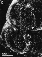 issued from : A. Ianora, A. Miralto & S. Vanucci in Mar Biol., 1992, 113. [p.403, Fig.2, C]. SEM micrographs. Female & Male: C, surface attachment structure (mass of fine setules arranged in three semicircles on a flattened area of the anterodorsal surface of the cephalosome). For the interpretation of this structure: see in Anomalocera patersoni.
|
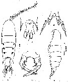 issued from : Z. Zheng, S. Li, S.J. Li & B. Chen in Marine planktonic copepods in Chinese waters. Shanghai Sc. Techn. Press, 1982 [p.87, Fig.49-1]. Female: a, habitus (dorsal); b, idem (lateral); c, urosome (ventral); d, P4; e, P5. Scale bars in mm.
|
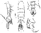 issued from : Z. Zheng, S. Li, S.J. Li & B. Chen in Marine planktonic copepods in Chinese waters. Shanghai Sc. Techn. Press, 1982 [p.88, Fig.49-2]. Scale bar in mm.
|
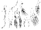 Issued from : G.S. Brady in Rep. Scient. Results Voy. Challenger, Zool., 1883, 8 (23). [Pl. XXXV, Figs.14-20]. Female: 14, habitus (dorsal); 15, A1; 16, Md; 17, P1; 18, P2; 19, P5; 20, urosome.
|
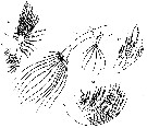 Issued from : G.S. Brady in Rep. Scient. Results Voy. Challenger, Zool., 1883, 8 (23). [Pl.XXXIV]. Female: 10, A2; 11, Mx1; 12, Mx2; 13, Mxp.
|
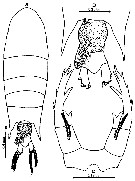 issued from : Mulyadi in Treubia, 2002, 32. [p.152, Fig.56]. Female (from Flores Sea): a, habitus (dorsal); b, posterodistal metasomal somite 5 and urosome (dorsal); c, P5. Nota:Cephalon with a projection over base of rostrum, separated from metasomal segment 1. Rostral ramus long and spiniform. Urosome 2-segmented; urosomal segment 1 asymmetrical, proximal right margin with 1 transparent setule, and 1 small and short medial spine-like process; left margin with medial swelling and 1 transparent setule proximally; urosomal segment 2 asymmetrical, right side longer. Caudal rami slightly asymmetrical, right ramus broader and longer. P5 similar that of P. macronyx, asymmetrical, right leg with 2 outer spinules and 3 distal spines of which innermost spine is longer and broader; inner margin of exopod at its distal half produced into 1 stout thick spinous process. Exopod of left leg produced into 1 stout spiniform process terminally, with 2 outer spines, distal one set in 3 spinules. Endopods left and right bifid at tip. In Krishnaswamy (1973) the spine on the right side of urosomite 1 of the female collected from Madras coast is lacking.
|
 issued from : P.P. Pillai in J. mar. biol. Ass. India, 1975, 17 (2). [p.136, Fig.3, c-e ]. Female (from Indian Ocean): c, urosome (dorsal); d, P5. Male: e, P5. Scale: Single bar: 0.3 mm; double bars: 0.05 mm.
|
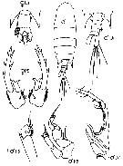 Issued from : J.M. Bradford-Grieve, E.L. Markhaseva, C.E.F. Rocha & B. Abiahy in South Atlantic Zooplankton, edit. D. Boltovskoy. 1999, Vol. 2, Copepoda; [p.1073, Fig. 7.403: Pontellopsis villosa]. Ur = urosome; r = right leg; l = left leg; r male A1 = male right antennula. Female characters (from key, p.960): - Urosome apparently of 1 somite in dorsal view. - Genital segment with large, hairy left side projection directed posteriorly, indistinctly separated from somite 2 in dorsal view. Male characters (from key, p.961): - Posterior prosome corners almost symmetrical, both points long, extending almost to posterior border of urosomal segment 3.
| | | | | Ref. compl.: | | | Sewell, 1948 (p.417, 419, 461); Krishnaswamy, 1953 (p.137); Demir, 1959 (p.176); Heinrich, 1960 a (p.43: fig.1, p.46: fig.2); Cervigon, 1962 (p.181, tables: abundance distribution); Björnberg, 1963 (p.61, Rem.); Heinrich, 1964 (p.87: fig.5, 91: fig.7); Shmeleva, 1965 b (p.1350, lengths-volume -weight relation); Mazza, 1966 (p.72); 1967 (p.367); Delalo, 1968 (p.138); Heinrich, 1969 (p.1457, fig.5); Bainbridge, 1972 (p.61, Appendix Table I: vertical distribution vs day/night); Champalbert & Gaudy, 1972 (p.159, respiration vs temperature); Apostolopoulou, 1972 (p.328, 369); Corral Estrada & Pereiro Muñoz, 1974 (tab.I); Heinrich, 1974 (p.43, fig.1); Weikert, 1975 (p.139, carte); Carter, 1977 (1978) (p.36); Vives, 1982 (p.295); Kovalev & Shmeleva, 1982 (p.85); Brenning, 1985 a (p.28, Table 2); Madhupratap & Haridas, 1986 (p.105, tab.1); Brenning, 1987 (p.30, Rem.); Lozano Soldevilla & al., 1988 (p.60); Heinrich, 1988 (p.89, fig.1); Echelman & Fishelson, 1990 a (tab.2); Suarez & al., 1990 (tab.2); Suarez & Gasca, 1991 (tab.2); Hernandez-Trujillo, 1991 (1993) (tab.I); Suarez, 1992 (App.1); Shih & Young, 1995 (p.72); Suarez-Morales & Gasca, 1997 (p.1525); Hure & Krsinic, 1998 (p.103); Suarez-Morales, 1998 (p.345, Table 1); Suarez-Morales & Gasca, 1998 a (p.111); Alvarez-Cadena & al., 1998 (tab.2, 3, 4); Dur & al., 2007 (p.197, Table IV); Morales-Ramirez & Suarez-Morales, 2008 (p.514); Schnack-Schiel & al., 2010 (p.2064, Table 2: E Atlantic subtropical/tropical); Mazzocchi & Di Capua, 2010 (p.427); Medellin-Mora & Navas S., 2010 (p.265, Tab. 2); Lidvanov & al., 2013 (p.290, Table 2, % composition); Bonecker & a., 2014 (p.445, Table II: frequency, horizontal & vertical distributions); Dias & al., 2015 (p.483, Table 2, abundance, biomass, production); Benedetti & al., 2016 (p.159, Table I, fig.1, functional characters); Marques-Rojas & Zoppi de Roa, 2017 (p.495, Table 1); El Arraj & al., 2017 (p.272, table 2); Belmonte, 2018 (p.273, Table I: Italian zones) | | | | NZ: | 14 | | |
|
Carte de distribution de Pontellopsis villosa par zones géographiques
|
| | | | | | | | | | | | 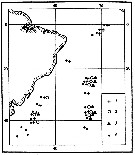 Issued from : A.K. Heinrich inTrudy Inst. Okeanol., 1974, 98 [p.44, Fig.1]. Issued from : A.K. Heinrich inTrudy Inst. Okeanol., 1974, 98 [p.44, Fig.1].
Distibution of three species of Pontellidae from SW Atlantic (Cruise of the R/V 'Akademik Kurchatov', 1971 and 1972).
Points = stations; cross = Labidocera acutifrons ; circle= Pontellopsis regalis ; triangle= Pontellopsis villosa . |
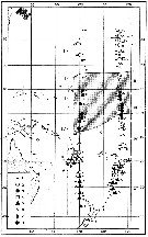 Issued from : A.K. Heinrich in Trudy Inst. Okeanol., 1960, 41. [p.43, Fig.1]. Issued from : A.K. Heinrich in Trudy Inst. Okeanol., 1960, 41. [p.43, Fig.1].
Distribution in the equatorial and tropical West Pacific of seven species of Pontellidae (Cruise of the R/V 'Vityaz', November 1957 to December 1958.
1: Stations; 2: Pontellopsis villosa; 3: Pontella fera; 4: Labidocera acutifrons; 5: Pontella whiteleggi; 6: Pontella tenuiremis; 7: Pontella chierchiae; 8: Labidocera acuta. |
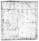 issued from : A.K. Heinrich in Zool. Zh., 1969, 48 (10) [p.1461, Fig.5]. issued from : A.K. Heinrich in Zool. Zh., 1969, 48 (10) [p.1461, Fig.5].
Distribution of two species of Pontellidae in the Pacific Ocean.
1: Pontellopsis villosa; 2: Pontella denticauda ( = Ivellopsis denticauda); 3: stations. |
 issued from : A.A. Shmeleva in Bull. Inst. Oceanogr., Monaco, 1965, 65 (n°1351). [Table 6: 36]. Pontellopsis vilosa (from South Adriatic). issued from : A.A. Shmeleva in Bull. Inst. Oceanogr., Monaco, 1965, 65 (n°1351). [Table 6: 36]. Pontellopsis vilosa (from South Adriatic).
Dimensions, volume and Weight wet. Means for 50-60 specimens. Volume and weight calculated by geometrical method. Assumed that the specific gravity of the Copepod body is equal to 1, then the volume will correspond to the weight. |
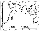 issued from : P. Pillai in J. mar. biol. Ass. India, 1975, 17 (2). [p.141, Fig.7]. issued from : P. Pillai in J. mar. biol. Ass. India, 1975, 17 (2). [p.141, Fig.7].
Occurrence of Pontellopsis villosa and P. armata collected from 32 stations in the Indian Ocean (See in: Hand Book to the International Zooplankton Collections, volume I, 1969). |
| | | | Loc: | | | South Africa, G. of Guinea, off Morocco-Mauritania, Canary Is., Portugal, Azores, Middle South Atlantic (off Mar del Plata), S Brazil, off Rio de Janeiro, Campos Basin, off Amazon, Caribbean Sea (Caribean Colombia, Bahia de Mochima (Venezuela), Cariaco Basin, Yucatan), E Costa Rica, G. of Mexico, Cuba, Florida, Georges Bank, Medit. (Alboran Sea, W Basin, Ligurian Sea, Tyrrhenian Sea, Strait of Messina, Adriatic Sea, Malta, Aegean Sea), G. of Aqaba, Red Sea, Natal, India ( Bombay coast, Madras coast), Burma coast, Andaman Sea, Indonesia-Malaysia (Sunda Strait, Cilacap Bay, Lombok Strait), Philippines, China Seas (East China Sea, South China Sea), Taiwan (SW), Japan, Tuamotu Is., Hawaii, Galapagos, W Baja California
Type locality: 30°32'N, 154°56'W. | | | | N: | 78 | | | | Lg.: | | | (16) F: 2,83-2,8; (46) F: 2,9-2,6; M: 2,5-2,3; (120) F: 2,7; (180) F: 2,8; (237) F: 3,0-2,75; M: 1,9; (256) F: 2,81-2,68; M: 2,48-2,05; (276) F: 2,62; 2,456; M: 2,576-2,536; (290) F: 1,95-2,45; M: 2,1; (795) F: 3; (1023) F: 2,38-2,47; M: 2,12-2,31; (1087) F: 2,55; (1115) F: 2,4-2,84; M: 2,11-2,14; {F: 1,95-3,00; M: 2,05-2,58} | | | | Rem.: | épipélagique. | | | Dernière mise à jour : 24/10/2022 | |
|
|
 Toute utilisation de ce site pour une publication sera mentionnée avec la référence suivante : Toute utilisation de ce site pour une publication sera mentionnée avec la référence suivante :
Razouls C., Desreumaux N., Kouwenberg J. et de Bovée F., 2005-2026. - Biodiversité des Copépodes planctoniques marins (morphologie, répartition géographique et données biologiques). Sorbonne Université, CNRS. Disponible sur http://copepodes.obs-banyuls.fr [Accédé le 07 février 2026] © copyright 2005-2026 Sorbonne Université, CNRS
|
|
 |
 |


