|
|
 |
Fiche d'espèce de Copépode |
|
|
Calanoida ( Ordre ) |
|
|
|
Clausocalanoidea ( Superfamille ) |
|
|
|
Scolecitrichidae ( Famille ) |
|
|
|
Scaphocalanus ( Genre ) |
|
|
| |
Scaphocalanus major (T. Scott, 1894) (F,M) | |
| | | | | | | Syn.: | Scolecithrix major T. Scott, 1894 b (p.52, Descr.F, figs.F); Sewell, 1948 (p.523); Giesbrecht & Schmeil, 1898 (p.47, Rem.F);
Scolecithrix ( Amallophora ) dubia var. similis T. Scott, 1894 b (p.56); Giesbrecht & Schmeil, 1898 (p.46, Rem.M); Vervoort, 1965 (p.64, Rem.); Sewell, 1948 (p.523, 564);
Scolecithrix similis : Esterly, 1905 (p.170, figs.M); in CalCOFI regional list (MDO, Nov. 2013; M. Ohman, comm. pers.);
Scaphocalanus similis : Brodsky, 1950 (1967) (p.257, figs.M);
Scolecithricella similis : Sewell, 1948 (p.516, 517, 557, 569);
? Amallophora media Sars, 1907 a (p.16);
? Scolecithrix gracilipes Farran, 1908 b (p.52, figs.F, Rem.);
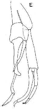
? Scaphocalanus medius : Sars, 1925 (p.173, figs.F); Rose, 1933 a (p.149, figs.F); C.B. Wilson, 1950 (p.328); Brodsky, 1950 (1967) (p.251, figs.F,M); Vaupel Klein, 1970 (p.19); Deevey & Brooks, 1977 (tab.2); Vives, 1982 (p.292); non S. major : Park, 1970 (p.476, 503, figs.F) | | | | Ref.: | | | A. Scott, 1909 (p.97, Rem.F); Sewell, 1948 (p.516, 546, 550); Vervoort, 1957 (p.110, Rem.); ? Tanaka, 1961 a (p.166, figs.F,M, Rem.); Vervoort, 1965 (p.63, Rem.); Minoda, 1971 (p.28); Bradford, 1973 (p.143); Park, 1982 (p.77, 108, figs.F, Rem.); Gardner & Szabo, 1982 (p.290, figs.F,M); Bradford & al., 1983 (p.93, 101, figs.F,M, Rem.); Boxshall & Halsey, 2004 (p.189, fig.M); Vives & Shmeleva, 2007 (p.772, figs.F,M, Rem.); Park & Ferrari, 2009 (p.143, Table 4, Appendix 1, biogeography from Southern Ocean) | 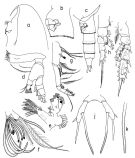 issued from T. Park in Biology of the Antarctic Seas XI, Antarct. Res. Ser, 1982, 34. [p.109, Fig.20]. Female: a, forehead; b, genital segment (lateral left side); c, last thoracic segment and urosome (lateral left side); d, Md; e, Mx1; f, distal part of Mx2; g, Mxp; h, P1 (anterior); i, P2 (posterior); j, P5 (posterior); k, distal part of inner spine of P5. Nota: Urosome about 37/100 length of prosome. All cephalosomal appendages similar to those of S. farrani
|
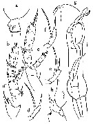
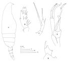 Issued from : J.M. Bradford, L. Haakonssen & J.B. Jillett in Mem. N.Z. oceanogr. Inst., 1983, 90. [p.102, Fig.60]. Female: A, habitus (lateral right side); B, endopod of P2; C, P5. Nota: The posterior matasomal corners of the Southwest Pacific are pointed not previously recorded by Tanaka (1961, 1982). The shape of the posterior metasome may be variable as it is more or less acutaly pointed according to the specimens of different stations. There seems to be some variation in the relative lengths of spines of P5, and the outer spine may be completely absent. Male: D, exopod 1 of P2; E, P5.
|
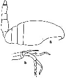 issued from : C.O. Esterly in Univ. Calif. Publs Zool., 1905, 2 (4). [p.170, Fig.31]. As Scolecithrix similis. Male (from San Diego Region): a, habitus (lateral); b, P5. Nota: Right A1 18-segmented, left 23-segmented. Last two thoracic segments fused. Genital segment short, 2nd long, twice the length of the 3rd, which is shorter than the 4th.
|
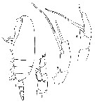 issued from : G.P. Farran in Fish. Ire. Sci. Invest., 1906, II [1908]. [Pl. VI, Figs.1-4]. As Scolecithrix gracilipes. Female (from W Ireland): 1, habitus (lateral); 2, P1; 3, P5; 4, P5 (abnormal).
|
 issued from : G.A. Boxshall & S.H. Halsey in The Ray Society, 2004, 166 [p.189, Fig.45, E]. After Vyshkvartzeva, 1989 b. Male: E, P5.
|
 issued from : O. Tanaka in Publ. Seto Mar. Biol. Lab., 1961, IX (1). [p.167, Fig.116]. Female (from Japan: Izu Region): a, last thoracic segment and genital segment (lateral, left side); b, P2; d', 1st segment of exopod of P2 (enlarged); c, P3; d-f, P5 (different specimens). Nota : Cephalothorax and abdomen 76 : 24. Last thoracic segment obtusely triangular on the posterior distal corner, but not pointed at the apex. Urosome 4-segmented ; urosomal segments and caudal rami in proportional lengths 42 : 25 : 23 : 9 : 21 = 100. The ventral margin distal to the genital opening furnished with hairs ; the genital, 2nd and 3rd segments fringed with fine teeth on the distal margin. Caudal rami about 2 times as long as wide. 1st segment of A1 furnished Endopod of A2 about 1.2 times as long as exopod.
Terminal spine of exopod of P2 with about 30 serrations..
Terminal spine of exopod of P3 with about 26 serrations
P5 2-segmented ; distal segment with 3 spines, the inner marginal spine about 2 times as long as the terminal one, and rather finely serrated on the outer border, the outer marginal spine is short, about ¼ the length of the terminal spine, and arises distal to the line of origin of the inner marginal spine.
Male: g, forehead (lateral); h, exopod of P2 (part.); i, P5.
Nota : Cephalothorax and urosome in proportional lengths 65 : 35.
Lateral margins of the head constricted much in front ; the frontal margin of the head obliquely rounded in lateral view.
Th5 and Th4 separate (clearly seen from the side).
Urosme 5-segmented ; urosomal segments and caudal rami in proportional lengths 9 :35 :23 :23 :1 :9 = 100.
Caudal rami about 1.4 times as long as wide (15 :11).
A1 19-segmented ( segments8 to 12 fused, 13-14 fused, 20-21 fused, and 24-25 fused).
Terminal spine of exopod of P2 with about 40 serrations.
P5 reaches back about to the distal end of the 2nd urosomal segment ; endopod of the left leg 2-segmented, and slightly inflated proximally.
|
 issued from : O. Tzanaka in Publ. Seto Mar. Biol. Lab., 1961, IX (1). [p.168]. Male: Proportional lengths of the segments od A1.
| | | | | Ref. compl.: | | | Grice & Hulsemann, 1965 (p.224); 1967 (p.16); Furuhashi, 1966 a (p.295, vertical distribution in Oyashio/Kuroshio transitional area, Table 8, 9); Roe, 1972 (p.277, tabl.1, tabl.2); 1972 b (p.532, Rem.); Harding, 1974 (p.141, tab.2, gut contents); Pipe & Coombs, 1980 (p.223, vertical occurrence); Vives, 1982 (p.292); Gowing & Wishner, 1986 (p.939, tab.3, gut contents); Lozano Soldevilla & al., 1988 (p.59); Gowing & Wishner, 1992 (tab.1); Shih & Young, 1995 (p.73); Razouls & al., 2000 (p.343, Appendix); Holmes, 2001 (p.59); Kuriyama & Nishida, 2006 (p.300: Tab;II; p.309: Tab.III, fig.7, 10, vertical distribution); Galbraith, 2009 (pers. comm.); in CalCOFI regional list (MDO, Nov. 2013; M. Ohman, pers. comm.); Sano & al., 2013 (p.11, Table 9, food habits). | | | | NZ: | 13 | | |
|
Carte de distribution de Scaphocalanus major par zones géographiques
|
| | | | | | | | | | | | 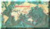 Carte de 1996 Carte de 1996 | |
| | | | Loc: | | | Antarct. (Indian, SE Pacif.), sub-Antarct. (Indian, SW & SE Pacif.), G. of Guinea, G. of Mexico, off Bermuda, off E Cape Cod, Canary Is., off W Ireland, Wyville Thomson Ridge, SW Indian, Indonesia-Malaysia, China Seas (South China Sea), Japan, Pacif. (NW & NE), British Columbia (rare), California, off W Mexico, New Zealand | | | | N: | 25 | | | | Lg.: | | | (5) F: 2,9; (9) F: 2,7-2,2; M: 2,2-1,8; (16) F: 2,6-2,4; M: 2,4; (47) F: 3; (76) F: 3,16-2,8; (108) F: 2,66; M: 2,76-2,55; (142) M: 2,6; ? (199) F: 2,66-2,28; M: 2,74-2,13; ? (208) F: 2,55-1,9; M: 2,8-2,6; (1000) F: 2,5 ± 0,2; M: 3,2 ± 0,1; {F: 1,90-3,16; M: 1,80-3,30} | | | | Rem.: | Méso-bathypélagique. Dans un trait vertical 2000-1000 m (Harding, 1974).
Sampling depth (Antarct., sub-Antarct.) : 0-1000-3000 m. 540-580 m (Pipe & Coombs, 1980 at 60°N, 07°W).
L'espèce présente une certaine variabilité.
Park (1982) suggère, en se fondant sur les dimensions, certaines synonymies entre S. major et S. medius . Dans ces conditions la répartition géographique de ces deux espèces demeure confuse, si elles ne sont pas synonymes comme l'estime Bradford.
Voir aussi les remarques en anglais | | | Dernière mise à jour : 19/01/2021 | |
|
|
 Toute utilisation de ce site pour une publication sera mentionnée avec la référence suivante : Toute utilisation de ce site pour une publication sera mentionnée avec la référence suivante :
Razouls C., Desreumaux N., Kouwenberg J. et de Bovée F., 2005-2026. - Biodiversité des Copépodes planctoniques marins (morphologie, répartition géographique et données biologiques). Sorbonne Université, CNRS. Disponible sur http://copepodes.obs-banyuls.fr [Accédé le 15 avril 2026] © copyright 2005-2026 Sorbonne Université, CNRS
|
|
|
|


;)
;)
;)
;)
;)
;)

;)
