|
|
 |
Fiche d'espèce de Copépode |
|
|
Calanoida ( Ordre ) |
|
|
|
Spinocalanoidea ( Superfamille ) |
|
|
|
Spinocalanidae ( Famille ) |
|
|
|
Spinocalanus ( Genre ) |
|
|
| |
Spinocalanus magnus Wolfenden, 1904 (F,M) | |
| | | | | | | Syn.: | Spinocalanus latifrons Sars, 1907 a (p.5);
Spinocalanus heterocaudatus Rose, 1937 (p.151, figs.F); Mazza, 1966 (p.70); no Rose, 1942 a (p.315, figs.M);
Spinocalanus pacificus Mori, 1942;
no S. magnus : Bradford, 1971 b (p.18, figs.F) | | | | Ref.: | | | Wolfenden, 1904 (p.118, Descr.F); Farran, 1905 (p.30, figs.F); Wolfenden, 1906 (p.42, figs.F, Descr.F); Farran, 1908 b (p.27); Wolfenden, 1911 (p.216, figs.F); With, 1915 (p.72, Rem.); Lysholm & Nordgaard, 1921 (p.11); Sars, 1925 (p.33, figs.F); Farran, 1926 (p.243); 1929 (p.208, 227, Rem.); Sewell, 1929 (p.95); Rose, 1933 a (p.85, figs.F); Jespersen, 1934 (p.52); Lysholm & al., 1945 (p.11); Vervoort, 1946 (p.150, Rem.); Davis, 1949 (p.22, figs.F); Brodsky, 1950 (1967) (p.125, figs. F); Farran & Vervoort, 1951 g (n°39, p.3, figs.F); Vervoort, 1951 (p.71, Rem.); Tanaka, 1956 c (p.391, figs.F,M); Vervoort, 1957 (p.41, Rem.); Harding, 1966 (p.33, figs.M, juv.F); Grice & Hulsemann, 1967 (p.21, Rem.M); Park, 1970 (p.475, 491, Rem.); Minoda, 1971 (p.21); Grice, 1971 (p.275, 280, figs.F); Vidal, 1971 a (p.18, 23, 119, figs.F,M, Rem.M); Damkaer, 1975 (p.26, figs.F, M, Rem.); Brodsky & al., 1983 ( p.257, figs.F,M, Rem.); Bradford-Grieve, 1994 (p.104, figs.F,M); Lapernat, 1999 (p.27, 55, Rem.); Bradford-Grieve & al., 1999 (p.878, 914, figs.F,M); Boxshall & Halsey, 2004 (p.197: fig.M); Vives & Shmeleva, 2007 (p.836, figs.F,M, Rem.); Bode & al., 2017 (p.600, Table I, III, fig. 2, 3, 4, morphology vs genetic). | 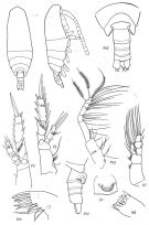 issued from : Brodsky K.A., Vyshkvartseva N.V., Kos M.S. & Markhaseva E.L. in Opred. Faune SSSR, 1983, 135. [p.258, Fig.118]. Female. Gnb = first inner lobe of Mx1; Gn = genital somite (ventral view).
|
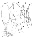 issued from : O. Tanaka in Publ. Seto Mar. Biol. Lab., V (3). [p.392, Fig.15]. Female: a, habitus (dorsal); b, head (left lateral view); c, last thoracic segment and urosome; (left side); d, P1; e, P2. Male: f, last thoracic segment with P5 and urosome (left lateral view); g, P5.
|
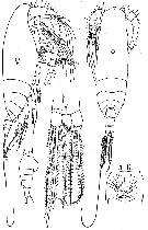
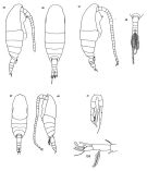 issued from : D.M. Damkaer in NOAA Technical Report NMFS CIRC-391, Seattle, 1975. [p.27, Fig.35-41; p.63, Fig.150]. Female: 35, habitus (right lateral side); 36, idem (dorsal); 37, idem (dufferent specimen); 38, distal part of prosome and urosome (dorsal); 150, terminal segments of A1. Male: 39, habitus (dorsal); 40, idem (left lateral side); 41, P5.
|
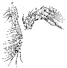
 issued from : G.D. Grice in Cah. Biol. Mar., 1971, XII. [p.278, Fig.3 G-H]. Female (from Medit.): G, habitus (lateral right side); H, Mxp.
|
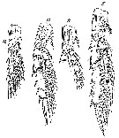
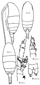 Issued from : K.A. Brodskii in Calanoida of the Far Eastern Seas and Polar Basin of the USSR. Opred. Fauna SSSR, 1950, 35 (Israel Program for Scientific Translations, Jerusalem, 1967) [p.125, Fig.43]. Female (from NP: NW Pacif; Ar: Arctic): habitus (dorsal and lateral left side); S1, P1; S3, P3; CDo, anal segment and caudal rami (dorsal). Nota: Caudal rami asymmetrical (left ramus longer than right ramus).
|
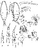 issued from : G.P. Farran in Ann. Rep. Fish. Brch., Ireland, 1902-1903, II, App. 2, 1905. [Plate III, Figs.1-12]. Female (from W Ireland): 1-2, habitus (dorsal and lateral, respectively); 3, proximal portion of A1; 4, A2; 5, Md; 6, Mx1; 7, Mx2; 8, Mxp; 9-111, P1 to P3; 12, basal segment of P4.
|
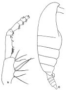
 issued from : M. Rose in Annls Inst. océanogr., Monaco, n. ser., 1937, XVII, 2. [p.152, Fig.1]. As Spinocalanus heterocaudatus. Female (from Alger Bay, Algeria): habitus (lateral and dorsal, respectively); Abd, urosome; A. G., genital segment (ventral); F, caudal rami. Nota: corners of the last thoracic segment reaching end of the genital segment. asymmetrical setae of the caudal rami very asymmetrical and curved. Endopodite of P1 with 5 setae.
|
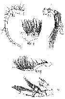 issued from : M. Rose in Annls Inst. océanogr., Monaco, n. ser., 1937, XVII, 2. [p.153, Fig.2]. As Spinocalanus heterocaudatus. Female: A1; A2; Mx2 (as Mxp1); Mxp (as Mxp2); P4.
|
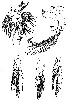 issued from : M. Rose in Annls Inst. océanogr., Monaco, n. ser., 1937, XVII, 2. [p.155, Fig.3]. As Spinocalanus heterocaudatus. Female: Mx1 (as Mx); Md; P1; P2; P3.
|
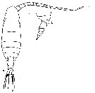 issued from : R.N. Wolfenden in Die Marinen Copepoden der Deutschen Südpolar-Expedition 1901-1903, 1911. [217, Fig.8]. Female: a, habitus (dorsal); b, posterior part cephalothorax and urosome (lateral).
|
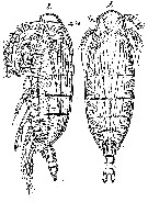 Issued from : G.O. Sars in Résult. Camp. Scient. Prince Albert I, 69, pls.1-127 (1924). [Pl.IX, figs. 8-9]. Female: 8, habitus (lateral); 9, idem (dorsal).
|
 Issued from : G.O. Sars in Résult. Camp. Scient. Prince Albert I, 69, pls.1-127 (1924). [Pl.IX, figs. 10-11]. Female: 10, A2; 11, Mxp.
|
 Issued from : G.O. Sars in Résult. Camp. Scient. Prince Albert I, 69, pls.1-127 (1924). [Pl.IX, figs. 12-15]. Female: 12, P1; 13, P2; 14, P3 (exopod inciomplete); 15, P4.
|
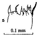 issued from : P.E. Lapernat & C. Razouls in Vie Milieu, 2002, 52 (1). [p.28, Pl. VI, fig.5]. Masticatory edge of Md gnathobase female (from off Malta, Mediterranean Sea). Nota: Itoh's index: 498.1 (number of teeth : 9).
|
 issued from : P.E. Lapernat & C. Razouls in Vie Milieu, 2002, 52 (1). [p.20, Pl. I, fig.5]. Masticatory edge of Md gnathobase female (from off Malta, Mediterranean Sea).
|
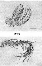 Issued from : M. Sano, K. Maki, Y. Nishibe, T. Nagata & S. Nishida in Progr. Oceanogr., 2013, 110. [p.19, Figs. 8 h, 9 h]. Second maxilla (Mx2) and maxilliped (Mxp) for Spinocalanus magnus from Sagami Bay (Japan) in April 2009. Scale bars: Mx2 & Mxp = 100 µm. Compare appendage structure and setal armement with Euchirella rostrata, Undeuchaeta major, Chirundina streetsii, Scaphocalanus echinatus, Scottocalanus helenae, Scottocalanus securifrons, Pleuromamma xiphias and calanus sinicus for interpreted particles catching.
| | | | | Ref. compl.: | | | Pearson, 1906 (p.10, Rem.); Damas & Koefoed, 1907 (p.400, tab.II); Hardy & Gunther, 1935 (1936) (p.157, Rem.); Massuti Alzamora, 1942 (p.110); Sewell, 1948 (p.347, 508, 555, 566, 569); C.B. Wilson, 1950 (p.341); Østvedt, 1955 (p.14: Table 3, p.59); Fagetti, 1962 (p.17); Grice, 1962 a (p.101); M.W. Johnson, 1963 (p.89, Table 1, 2); V.N. Greze, 1963 a (tabl.2); Harding, 1966 (p.17, 65, 66, 71); Sewell, 1948 (p.347, 495, 508, 513, 526, 545, 548, 555, 566, 568, 574); Grice, 1963 a (p.495); Unterüberbacher, 1964 (p.20); De Decker & Mombeck, 1964 (p.14); Grice & Hulsemann, 1965 (p.223); Furuhashi, 1966 a (p.295, vertical distribution in Kuroshio region, Table 10); Fleminger, 1967 a (tabl.1); Dunbar & Harding, 1968 (p.319, 320); Vinogradov, 1968 (1970) (p.266, 258); Roe, 1972 (p.277, tabl.1, tabl.2); 1972 a (p.329); Björnberg, 1973 (p.322, 389); Harding, 1974 (p.141, Table 2, 3, gut contents); Vives & al., 1975 (tab.II, XII); Deevey & Brooks, 1977 (p.256, tab.2, Station "S"); Vives, 1982 (p.290); Kovalev & Schmeleva, 1982 (p.83); Roe, 1984 (p.356); Scotto di Carlo & al., 1984 (1042); Guangshan & Honglin, 1984 (p.118, tab.); Brenning, 1985 a (p.28, Table 2); Hopkins, 1985 (p.197, Table 1, gut contents); Lozano Soldevilla & al., 1988 (p.58); Scotto di Carlo & al., 1991 (p.270); Hattori, 1991 (tab.1, Appendix); Mumm, 1993 (tab.1, fig.2); Ward & al., 1995 (p.195, Table 2); Shih & Young, 1995 (p.74); Hure & Krsinic, 1998 (p.41, 100); Padmavati & al., 1998 (p.349); Hure & Krsinic, 1998 (p.41, 100); Razouls & al., 2000 (p.343, tab. 5, Appendix); Lapernat & Razouls, 2001 (tab.1); Weikert & al., 2001 (p.229, fig.7, Rem.); Holmes, 2001 (p.42); Yamaguchi & al., 2002 (p.1007, tab.1); Vukanic, 2003 (139, tab.1); Ikeda & al., 2006 (p.1791, Table 2); Gaard & al., 2008 (p.59, Table1, N Atlantic Mid-Ridge); Darnis & al., 2008 (p.994, Table 1); Galbraith, 2009 (pers. comm.); Park & Ferrari, 2009 (p.143, Table 4, Appendix 1, biogeography); Homma & Yamaguchi, 2010 (p.965, Table 2); Schnack-Schiel & al., 2010 (p.2064, Table 2: E Atlantic subtropical/tropical); Mazzocchi & Di Capua, 2010 (p.427); Medellin-Mora & Navas S., 2010 (p.265, Tab. 2); Homma & al., 2011 (p.29, Table 2, abundance, feeding pattern: suspension feeders); in CalCOFI regional list (MDO, Nov. 2013; M. Ohman, comm. pers.); Sano & al., 2013 (p.11, Rem.: p.21, Table 2, 5, 6, A1, figs.5, 8, 9, feeding habits, vertical distribution); Zaafa & al., 2014 (p.67, Table I, occurrence); Belmonte, 2018 (p.273, Table I: Italian zones) | | | | NZ: | 20 | | |
|
Carte de distribution de Spinocalanus magnus par zones géographiques
|
| | | | | | | | | | | | | | | | | | 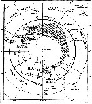 issued from : W. Vervoort in B.A.N.Z. Antarctic Reseach Expedition, Reports - Ser. B, Vol. III, 1957 [Fig.11] issued from : W. Vervoort in B.A.N.Z. Antarctic Reseach Expedition, Reports - Ser. B, Vol. III, 1957 [Fig.11]
Chart showing the geographical distribution (white circle) in the seas surrounding the Antarctic continent.
Nota: In this chart the area frequented by whaling vessels has been hatched. The Antarctic circle (66°.5 S) has been drawn as a broken line. The numbers I to VI refer to the sectors into which the Antarctic seas are divided according to Mackintosh (1942) (after Vervoort, 1951). |
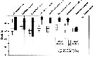 Issued from : M. Sano, K. Maki, Y. Nishibe, T. Nagata & S. Nishida in Progr. Oceanogr., 2013, 110. [p.20, Fig.10]. Issued from : M. Sano, K. Maki, Y. Nishibe, T. Nagata & S. Nishida in Progr. Oceanogr., 2013, 110. [p.20, Fig.10].
Vertical distribution of eight copepod species across 0-1000 m in Sagami bay in 21-27 April 2009.
White and black boxes denote daytime and nighttime distributions. Scaphocalanus echinatus/em> is taken from Kuriyama & Nishida (2006).
Plankton and water samples collected from a fixed station (35°00'N, 139°20'E) using MTD horizontal closing nets (0.33 mm mesh aperture) at 15 depths (0, 50, 95, 100, 150, 195, 200, 295, 300, 395, 400, 500, 600, 800, 1000 m) during both day and night.
Atomic C:N ratio (mean ± SD) = 14.0 ±1.5, n = 3. |
| | | | Loc: | | | Antarct. (Croker Passage, Peninsula, Weddell Sea, SW Atlant., Indian, SW Pacif., Ross Sea), sub-Antarct. (South Georgia, Indian, SE Pacif.), South Africa (E), Namibia, SE Atlant., Mauritania, Canary Is., off W Cabo Finisterre, Bay of Biscay, Azores, Caribbean Sea, Caribbean Colombia, G. of Mexico, Sargasso Sea, Station "S" (32°10'N, 64°30'W), off E Cape Cod, Davis Strait, Baffin Bay, Arct. (Fletcher's Ice Is., SE Beaufort Sea), Faroe Is., off W Ireland, Porcupine Bank, Norway Sea, North Sea, Ibero-moroccan Bay, Medit. (M'Diq, Alboran Sea, W Basin, Ligurian Sea, Tyrrhenian Sea, off Malta, S Adriatic Sea, Ionian Sea, E Basin), Arabian Sea, off S Laccadive Is., Indian, Indonesia-Malaysia, China Seas (East China Sea, South China Sea), Japan, Sagami Bay, off Sanriku, Pacif. (NW), Bering Sea, Aleutian Basin, Arct. (polar Basin), Chukchi Sea, Pacif. (W equatorial), Pacif. (equatorial), off British Columbia, off California, Tuamotu Is., off Galapagos, Chile | | | | N: | 95 | | | | Lg.: | | | (1) F: 2,4; (7) F: 2,8; (10) F: 2,8-2,6; (13) F: 3,1-1,87; M: 2,43-1,8; (22) F: 3-2,6; (25) F: 2,75-2,35; (28) F: 2,45-1,88; (31) F: 2,65-2,4; (35) F: 2,76-2,4; (38)?F : 2,67-2,58; (55) F: 2,5; M: 2,13; (58) F: 2,9; (59) F: 2,9-2,2; (67) M: 2; (88) F: 2,44-2,16; (199) F: 2,96-2,28; M: 2,43; (208) F: 2,5; (229) F: 2,75; (238) F: 2,8-2,75; (340) F: 2,35; 2,1; (432) F: 2,95-2,87; (545) F: 2; (1252) F: 2,20-2,55; {F: 1,87-3,10; M: 1,80-2,43}.
(1252) F: Pr/Ur = 2,8-3,8. | | | | Rem.: | épi-mésopélagique à abyssale.
Sampling depth (Antarct., sub-Antarct.) : 400-1000 m.
Brodsky & al. (1983, p.260) estiment qu'il n'y a pas de synonymie entre les mâles de cette espèce et ceux de S. spinipes.
Voir aussi les remarques en anglais | | | Dernière mise à jour : 28/03/2020 | |
|
|
 Toute utilisation de ce site pour une publication sera mentionnée avec la référence suivante : Toute utilisation de ce site pour une publication sera mentionnée avec la référence suivante :
Razouls C., Desreumaux N., Kouwenberg J. et de Bovée F., 2005-2026. - Biodiversité des Copépodes planctoniques marins (morphologie, répartition géographique et données biologiques). Sorbonne Université, CNRS. Disponible sur http://copepodes.obs-banyuls.fr [Accédé le 29 mars 2026] © copyright 2005-2026 Sorbonne Université, CNRS
|
|
|
|


;)
;)
;)
;)
;)
;)
;)
;)
;)
;)
;)
;)
;)
;)
;)
;)

;)
;)
