|
|
 |
Fiche d'espèce de Copépode |
|
|
Calanoida ( Ordre ) |
|
|
|
Clausocalanoidea ( Superfamille ) |
|
|
|
Aetideidae ( Famille ) |
|
|
|
Euchirella ( Genre ) |
|
|
| |
Euchirella pseudotruncata Park, 1975 (F,M) | |
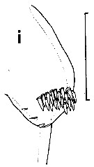
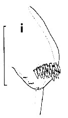
| | | | | | | Syn.: | Euchirella splendens (M) : Grice, 1969 a (p.453, figs.M) | | | | Ref.: | | | Park, 1975 (p.291, figs.F,M); 1976 a (p.115, figs.F,M); Bradford & Jillett, 1980 (p.33); Vaupel Klein, 1980 (p.153); 1984 a (p.43, Table II: characters, fig.F); Markhaseva, 1996 (p.163, figs.F,M) | 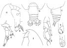 issued from : T. Park in Contr. Mar. Sci., 1976, 20. [p.115, Fig.6]. Female: a, forehead (lateral); b, c, posterior part of metasome and urosome (dorsal, lateral, respectively); d, A2; e, coxa of fourth leg (posterior). Male: f, forehead (lateral); g, fifth pair of legs (anterior); h, distal part of left fifth leg (anterior); i, idem (posterior).
|
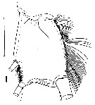
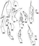 issued from : G.D. Grice in Bull. mar. Sc., 1969, 19 (2). [p.450, Figs.35-48]. As Euchirella splendens. Male (from Carubbean Sza & G. of mexico): 35, habitus (lateral left side); 36, forehead (lateral); 37, A2: 38, Mx1; 39, Mx2; 40, Mxp; 41, P1; 42, P2; 43, P3; 44, P4: 45, P5; 46, distal end of left P5 (enlarged); 47, P5 (other side); 48, distal end of left P5 (other side, enlarged). Nota: Presence of a prominent spinous process on the proximal end of endopod of right P5. Terminal portion of left P5 with a small spinelike point and numerous small rounded knobs.
|
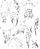 issued from : T. Park in Crustaceana, 1975, 28 (3). [p.292, Fig.1]. Female (from Gulf of Mexico): A-B, habitus (dorsal and lateral, respectively); C-D, posterior par of body (dorsal and lateral, respectively); E, forehead (lateral); F, A2; G, P1 (anterior); H, P2 (anterior); I, basipod of P4 (posterior).
|
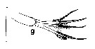
 issued from : T. Park in Crustaceana, 1975, 28 (3). [p.293, Fig.2]. Male: A-B, habitus (dorsal and lateral, respectively); C, P5 (anterior); D, distal part of left P5 (anterior); E, idem (posterior). Nota: P5 consisting of a small, uniramose left leg and a well developed, biramose right leg; right basipod reaching about middle of 1st exopodal segment; right endopod unsegmented, tapering into a curved spiniform process, with a prominent tooth-like process close to proximal end; right exopod 2-segmented, proximal segment with a large tooth-like process at a level immediately posterior to the tooth-like process on endopod; distal segment tapering into a curved spiniform process, with its internal margin finely serrated. In left leg of P5 endopod lacking; exopod 3-segmented, last segment small with 3 or 4 small tooth-like processes distally, and together with distal part of 2nd segment (immovable finger), forming a small chela; immovable finger of the chela slightly longer than movable finger (3rd segment).
|
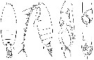
 issued from : J.C. von Vaupel Klein in Crustaceana, Supplt 9, Studies on Copepoda, III, 1984. [p.76, Fig.14, i]. Female: i, arrangement of spinules on the lateral tubercle of ebdopod of P1 (note: spinules approximately equal size, and arranged in a close group). Scale bar 0.067 mm.
|
 issued from : J.C. von Vaupel Klein in Crustaceana, Supplt 9, Studies on Copepoda, III, 1984. [p.78, Fig.15, l]. Female: i, fusion of exopodal segments 1 and 2 of P1 (left appendage, posterior view). Nota: Only very faint indications of the former suture are occasionally found in individual specimens of most species of Euchirella, and then mainly on the anterior face of the combined segment. Scale bar 0.1 mm.
|
 issued from : J.C. von Vaupel Klein in Crustaceana, Supplt 9, Studies on Copepoda, III, 1984. [p.69, Fig.9, g]. Female: g, complement of terminal setae on the 2nd inner lobe of basipodal segment 1 of right Mx1 (details of right appendages in anterior view; to the posterior side 2 relatively stout setae are always present, which are combined bipectinate and spinulose; usual Euchirella differentiation in 2 subequal anterior setae: the distal seta bipectinate/spinulose, the proximal one spinulose only). Scale bar: 0.2 mm.
|
 issued from : J.C. von Vaupel Klein in Crustaceana, Supplt 9, Studies on Copepoda, III, 1984. [p.76, Fig.14, g]. Female: g, arrangement of spinules on the lateral tubercle of endopod of right P1 . Scale bar: 0.067 mm. Nota: Spinules approximately equal size, and arranged in a close group.
|
 Euchirella pseudotruncata Euchirella pseudotruncata female: 1 - Genital segment slightly asymmetrical. 2 - Crest absent. 3 - Genital segment with left lateral swelled (dorsal view). 4 - Coxopodite of P4 with 1 spine. 5 - Posterior border of genital segment without projection on the right. The projection of left lateral margin of segment, occupying nearly all segment' length (dorsal view). 6 - Genital segment without ear-like projections.
|
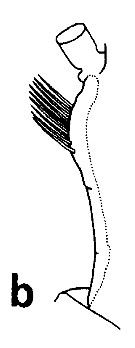
 Euchirella pseudotruncata Euchirella pseudotruncata male: 1 - P5 uniramous on left P5 and large biramous on right P5. Exopodal segment 2 and endopod of right P5 elongated and sharpened in their distal parts forming tongs. Endopod of right P5 significantly exceeding distal border of exopodal segment 1 of right P5. 2 - Crest absent. 3 - No spine present near place wherefrom exopod of right P5 begins. 4 - Left P5 without rudimentary endopod. When closed tongs of exopod of left P5 compact, rounded, clavate-like. 5 - Proximal part of exopodal segment 1 of right P5 with 1 big tooth. 6 - Proximal part of endopodal segment 1 of right P5 with robust tooth.
|
 issued from : J.C. von Vaupel Klein in Crustaceana, Supplt 9, Studies on Copepoda, III, 1984. [p.66, Fig.7, b]. Euchirella pseudotruncata a, A2 arrangements of spinules and hairs on the posterior edge of endopodite 2+3 (right appendage of medial view ): distal spinules.
|
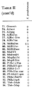
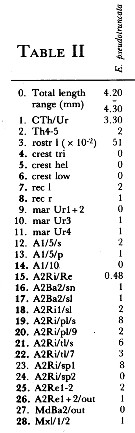 Issued from : J.C. von Vaupel Klein in Crustaceana, (Supplement) 9, 1984. [p.93, Table II]. Euchirella pseudotruncata Female: Datamatrix stating observed states of characters from Table I (p.87-90) presently examined; nos. refer to the input nos. used in Table I (see to the family Aetideidae).
|
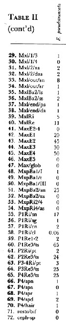 Issued from : J.C. von Vaupel Klein in Crustaceana, (Supplement) 9, 1984. [p.94, Table II (cont' d) ]. Euchirella pesudotruncata Female: Datamatrix stating observed states of characters from Table I (p.87-90) presently examined; nos. refer to the input nos. used in Table I (see to the family Aetideidae).
|
 Issued from : J.C. von Vaupel Klein in Crustaceana, (Supplement) 9, 1984. [p.95, Table II (cont' d) ]. Euchirella pseudotruncata Female: Datamatrix stating observed states of characters from Table I (p.87-90) presently examined; nos. refer to the input nos. used in Table I (see to the family Aetideidae).
| | | | | Ref. compl.: | | | Suarez-Morales & Gasca, 1998 a (p107); Lopez-Salgado & al., 2000 (tab.1); Lapernat, 2000 (tabl.3, 4) | | | | NZ: | 2 | | |
|
Carte de distribution de Euchirella pseudotruncata par zones géographiques
|
| | | | Loc: | | | Gulf of Mexico, Caribbean Sea, Sargasso Sea, off NE Cape Verde Is.
Type locality: Gulf of Mexico. | | | | N: | 6 | | | | Lg.: | | | (225) M: 3,60-3,48; (235) F: 4,66-4; M: 3,8-3,32; (236) F: 4,66-4,3; M: 3,8-3,64; (1257) F: 4,2-4,3; {F: 4,00-4,66; M: 3,32-3,80} | | | | Rem.: | épi-bathypélagique.
Voir aussi les remarques en anglais | | | Dernière mise à jour : 22/02/2021 | |
|
|
 Toute utilisation de ce site pour une publication sera mentionnée avec la référence suivante : Toute utilisation de ce site pour une publication sera mentionnée avec la référence suivante :
Razouls C., Desreumaux N., Kouwenberg J. et de Bovée F., 2005-2026. - Biodiversité des Copépodes planctoniques marins (morphologie, répartition géographique et données biologiques). Sorbonne Université, CNRS. Disponible sur http://copepodes.obs-banyuls.fr [Accédé le 08 janvier 2026] © copyright 2005-2026 Sorbonne Université, CNRS
|
|
|
|


;)
;)
;)
;)
;)
;)
;)
;)
;)
;)
;)
;)
