|
|
 |
Fiche d'espèce de Copépode |
|
|
Calanoida ( Ordre ) |
|
|
|
Arietelloidea ( Superfamille ) |
|
|
|
Arietellidae ( Famille ) |
|
|
|
Paramisophria ( Genre ) |
|
|
| |
Paramisophria aegypti Zagami, Bonanzinga & Costanzo, 2019 (F,M) | |
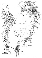
| | | | | | | Ref.: | | | Zagami & al., 2019 (p.'273, Descr.F, M, figs. F, M, Rem.) |  Issued from : G. Zagami, V. Bonanzingua & G. Costanzo in J. Nat. Hist., 2019, 53 (5-6). [p.276, Fig.2]. Male (from 27.4337°N, 34.1510°E): a, habitus (dorsal); b, left A1; c, right A1. Nota male: - Body symmetrical. - Rostrum with a pair of filaments on tip (identical to P. mediterranea. - Cepghalosome and 1st pedigerous somite separate, 4th and 5th somites completely fused, with dorso-lateral process and rounded postero-lateral lobe. Shape of pediger 5 with its process and lobe identical to P. sinjinensis. - Urosome 5-segmented. - Caudal rami symmetrical, about 1.8 times longer than wide, armed with 7 setae. - A1 strongly asymmetrical; left longer than rigght one. Left A1 18-segmented with geniculation between segments 17 (XX) and 18 (XXI-XXVIII). Right A1 21-segmented.
|
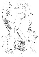
 Issued from : G. Zagami, V. Bonanzingua & G. Costanzo in J. Nat. Hist., 2019, 53 (5-6). [p.277, Fig.3]. Male: a, A2; b, Md; c, Mx1; d, Mx2; e, Mxp. A2 with coxa without setae; bais with inner terminal seta; endopod 2-segmented; exopod7-segmented indistinctly, 1st and 2nd segments without setae, 3rd to5th segments with 1, 2, 1 setae, respectively, 6th segment with 1 medial ,seta, 7th segment bearing 2 distal setae. - Md: gnathobase bearing 4 sharp teeth, 2 short processes and 2 distal patches of spinules. Palp with basis bearing a row of setules on outer margin; endopod reduced with 2 setae; exopod 5-segmented, 2nd segment longest, armature formula: 1, 1, 1, 1, 2. Mx1: praecoxal arthrite bearing 4 stout soines plus 1 reduced spine-like process; coxal endite without setae, coxal epipodite bearing 8 setae; basis without endites; endopod 1-segmented with 2 unequal setae, distally; exopod incorporated into basis, with 3 distal long setae. Mx2: praecoxa and coxa partially fused to form syncoxa. Proximal praecoxal endite with 1 seta, distal enditewith 2 setae. Coxal endites each with 2 setae. Basis bearing 1 stout spine on medial margin. Endopod with 8 stout pectinate setae. Mxp: syncoxa bearing 1 medial and 2 subdistal setae. Basis partially fused to 1st endopodal segment, ornamented with patch of spinules and armed with 2 basal setae and 1 endopodal seta. Endopod 5-segmented , armature formula 4, 4, 3, 3, 4.
|
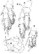
 Issued from : G. Zagami, V. Bonanzingua & G. Costanzo in J. Nat. Hist., 2019, 53 (5-6). [p.279, Fig.4]. Male: a, P1; b, P2; c, P4; d, P5. P5 asymmetrical. Coxae and intercoxal sclerite unarmed. Right leg uniramous; basis with 1 short seta about mid-length and inner margin produced into prominent lobe; exopod 3-segmented, 1st segment with distal outer margin produced into elongate lobe bearing terminal seta; 2nd segment with 1 subdistal seta on outer margin; 3rd segment elongated with 2 unequal setae and 1 long and stout terminal process completely incorporated into segment. Ledt leg biramous; basis bearing 1 stout plumose seta; endopod short reaching tip of 1st exopodal segment; exopd 3-segmented, 1st segment with terminal seta; 2nd segment with subdistal seta on outer margin; 3rd segment very reduced with 2 equal outer setules, shorter and thinner than right counterpart ones, and 1 long and stout inner process completely incorporated into segment.
|
 Issued from : G. Zagami, V. Bonanzingua & G. Costanzo in J. Nat. Hist., 2019, 53 (5-6). [p.278]. Male and female: Spine (Roman numerals) and seta (Arabic numerals) formula of the swimming legs P1 to P4.
|
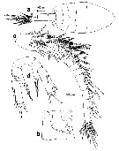
 Issued from : G. Zagami, V. Bonanzingua & G. Costanzo in J. Nat. Hist., 2019, 53 (5-6). [p.280, Fig.5]. Female (paratype): a, habitus (dorsal); b, genital double-somite (ventral view; copulatory pore indicated by arrow; c, left A1; d, P5. Nota female: - Body shape similar to male. - Urosome 4-segmented. - Oral parts and remaining legs as described in male. - Genital double-somite asymmetrical, with a pair of ventrolateral gonopores; single copulatory pore located on right side, nearly the middle part of somite. - Left A1 21-segmented. Right A1 as in male except for absence of aesthetasc on segment 2 (IV). - P5 asymmetrical. Coxa and intercoxal sclerite fused to form unarmed segment. Basis end endopod completely fused, outer seta on basis more developed and plumose in left leg than naked right one; endopodal lobe with long plumose seta and acute process terminally. Exopod 1-segmented,elongated, about 3.4 times longer than wide, bearing 3 short spines along outer margin, 1 long distal spine, and 1 short subdistal spine on inner margin; distal spine about 12.8 times longer than subdistal spine; stout process between terminal spine and subdistal inner soine.
| | | | | NZ: | 1 | | |
|
Carte de distribution de Paramisophria aegypti par zones géographiques
|
| | | | Loc: | | | N Red Sea (Ras Mohamed)
Type locality: 27.4337° N, 34.1510° E. | | | | N: | 1 | | | | Lg.: | | | (1234) F: 1,05- 1,09; M:: 1,00-1,03; {F: 1,05-1,09; M: 1,00-1,03} | | | | Rem.: | Collected from the sandy bottom of a fissure (3-5 m deep) connected with the sea. For Zagami & al. (2019, p.281) this species appears closely related to P. ammophila and P. platysoma, with which it shares the following combination of characters:
- 4th and 5th pedigerous somites completely fused wit dorsolateral processes and rounded postero-lateral lobes;
- Male left P5 with short endopod.- Both 3rd exopodal segments of the male P5 carry a long a stout terminal process completely incorporated into segment.
This combination of characters is unique among Paramisophria species. | | | Dernière mise à jour : 19/06/2023 | |
|
|
 Toute utilisation de ce site pour une publication sera mentionnée avec la référence suivante : Toute utilisation de ce site pour une publication sera mentionnée avec la référence suivante :
Razouls C., Desreumaux N., Kouwenberg J. et de Bovée F., 2005-2026. - Biodiversité des Copépodes planctoniques marins (morphologie, répartition géographique et données biologiques). Sorbonne Université, CNRS. Disponible sur http://copepodes.obs-banyuls.fr [Accédé le 08 février 2026] © copyright 2005-2026 Sorbonne Université, CNRS
|
|
 |
 |


