|
|
 |
Fiche d'espèce de Copépode |
|
|
Calanoida ( Ordre ) |
|
|
|
Diaptomoidea ( Superfamille ) |
|
|
|
Candaciidae ( Famille ) |
|
|
|
Candacia ( Genre ) |
|
|
| |
Candacia cheirura Cleve, 1904 (F,M) | |
| | | | | | | Syn.: | Candacia chirura : Fleminger & Bowman, 1956 (p.336, fig.F); Unterüberbacher, 1964 (p.31, figs.F,M)) | | | | Ref.: | | | Cleve, 1904 a (p.196: as chirura, p.198, F & M, Pl. I,II); Farran, 1929 (p.210, 273, figs.F,M, Rem.); Vervoort, 1957 (p.142, Rem.); Grice, 1963 (p.175, 182, 189); Unterüberbacher, 1964 (p.31, figs.F, M); Ramirez, 1971 (p.87); Bradford, 1972 (p.48, figs.F,M); Lawson, 1977 (p.71, tab.2, 3, 4, fig.5); Björnberg & al., 1981 (p.657, figs.F,M); Mazzocchi & al., 1995 (p.71, figs.F,M, Rem.); Bradford-Grieve & al., 1999 (p.885, 956, figs.F,M); Bradford-Grieve, 1999 b (p.166, figs.F,M, Rem., figs.182, 193); Park & Ferrari, 2009 (p.143, Table 3, Appendix 1, biogeography from Southern Ocean) | 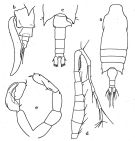 issued from : G.P. Farran in British Antarctic (\"Terra Nova\") Expedition, 1910. Natural History Reports. Zoology. Vol. VIII. Crustacea, 1929. [p.274, Fig.29]. Female: a, habitus (dorsal view); b, urosome (left lateral view). Male: c, distal part of prosome and urosome (dorsal view); d, clasping joints of right A1; e, fifth feet.
|
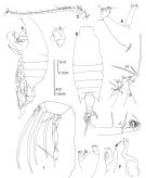 issued from : Bradford-Grieve J.M. in The Marine Fauna of New Zealand: Pelagic Calanoid Copepoda. National Institute of Water and Atmospheric Research (NIWA). NIWA Biodiversity Memoir, 111, 1999. [p.162, Fig.113]. Female: A, habitus (left lateral side); B, idem (dorsal); C, genital somite (left lateral side); D, A1; E, A2; F, Md (mandibular blade); G, Md (mandibular palp); H, Mx1; I, Mx2; J, Mxp.
|
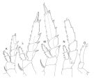 issued from : Bradford-Grieve J.M. in The Marine Fauna of New Zealand: Pelagic Calanoid Copepoda. National Institute of Water and Atmospheric Research (NIWA). NIWA Biodiversity Memoir, 111, 1999. [p163 (legend in Fig.113). Female: K, P1; L, P2; M, P3; N, P4; O, P5.
|
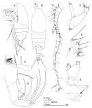 issued from : Bradford-Grieve J.M. in The Marine Fauna of New Zealand: Pelagic Calanoid Copepoda. National Institute of Water and Atmospheric Research (NIWA). NIWA Biodiversity Memoir, 111, 1999. [p.164, Fig.114]. Male: A, habitus (left lateral side); B, idem (dorsal); C, right A1; D, A2; E, Md; F, Mx1; G, Mx2; H, Mxp.
|
 issued from : Bradford-Grieve J.M. in The Marine Fauna of New Zealand: Pelagic Calanoid Copepoda. National Institute of Water and Atmospheric Research (NIWA). NIWA Biodiversity Memoir, 111, 1999. [p.165, (legend in Fig.114]. Male: I, P1; J, P2; K, P3; L, P4 (exopod segment 3 inner edge with 4 setae on one side and 5 on the other); M, P5 (L = left leg; R = right leg); N, terminal part of right P5.
|
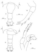 Issued from: M.G. Mazzocchi, G. Zagami, A. Ianora, L. Guglielmo & J. Hure in Atlas of Marine Zooplankton Straits of Magellan. Copepods. L. Guglielmo & A. Ianora (Eds.), 1995. [p.72, Fig.3.8.1]. Female: A, urosome (dorsal); B, P5. Nota: Proportional lengths of urosomites and furca 44 : 26 : 15 : 15 = 100. Male: C, urosome (dorsal); D, P5. Nota: Proportional lengths of urosomites and furca 18 : 24 : 21 : 13 : 10 : 14 = 100.
|
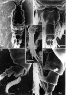 Issued from: M.G. Mazzocchi, G. Zagami, A. Ianora, L. Guglielmo & J. Hure in Atlas of Marine Zooplankton Straits of Magellan. Copepods. L. Guglielmo & A. Ianora (Eds.), 1995. [p.73, Fig.3.8.2]. Female (SEM preparation): A, habitus (sorsal); B, urosome (dorsal); C, urosome bearing spematophore (lateral right side); D, large protuberance on left side of 2nd urosomal somite; E, P5 Bars: A-D 0.100 mm; E 0.050 mm.
|
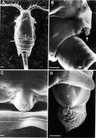 Issued from: M.G. Mazzocchi, G. Zagami, A. Ianora, L. Guglielmo & J. Hure in Atlas of Marine Zooplankton Straits of Magellan. Copepods. L. Guglielmo & A. Ianora (Eds.), 1995. [p.74, Fig.3.8.3]. Male (SEM preparation): A, habitus (dorsal); B, right corner of last thoracic and 1st urosomal somites; C, detail of flap-like projection on dorsal side of 1st thoracic somite (arrow in A); D, detail of acorn-like swelling on right corner of thorax (arrow in B). Bars: A 0.100 mm; B 0.050 mm; C-D 0.010 mm.
|
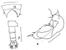 issued from : F.C. Ramirez in Revta Mus. La Plata, Seccion Zool., 1971, XI.[Lam.II, Figs.4, 7, 11]. Male (from off Patagonia): 4, right P5 (distal segments); 7, urosome (dorsal); 11, posterior part cephalothorax. Scale bars in mm: 0.4 (4, 7, 11).
|
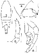 issued from : J.M. Bradford in Mem. N. Z. Oceonogr. Inst., 1972, 54. [p.49, Fig.13, (3-6). Female (from Kaikoura, New Zealand): 3, habitus (dorsal); 5, P5. Male: 4, prortion of last thoracic segment and genital segment (dorsal); 6, P5. Scale bars: 1 mm (3); 0.1 mm (4, 5, 6).
|
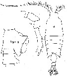 issued from : H.K. Unterüberbacher in Investl Rep. mar. Res. Lab. S.W. Afr., 1964, 11. [Pl.31]. As Candacia chirura. Female (from off Namibia): habitus (dorsal); P5, genital segment and urosomal segment 2 (lateral, right side).
|
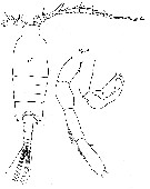 issued from : H.K. Unterüberbacher in Investl Rep. mar. Res. Lab. S.W. Afr., 1964, 11. [Pl.32]. As Candacia chirura. Male: habitus (dorsal), P5.
|
 issued from : A. Fleminger & T.E. Bowman in Proc. U.S. Natn. Mus. 1956, 106, No 3370. [p.335, Fig.2, n]. Female (after Farran, 1929): n, urosome (lateral, left side).
|
 Issued from : J.M. Bradford-Grieve, E.L. Markhaseva, C.E.F. Rocha & B. Abiahy in South Atlantic Zooplankton, edit. D. Boltovskoy. 1999, Vol. 2, Copepoda; [p.1064, Fig. 7.364: Candacia cheirura ]. Ur = urosome; r = right leg; l = left leg. Female characteristics (from key, p.956) : - In lateral view, ventral protrusion of urosomal segment 2 directed obliquely posteriad; terminal segment of P5 with 2 small, outer, spine-like points and 3 spine-like points distally, middle one of which is longest. - In dorsal view, protrusion on ventral surface or urosomal segment 2 not visible; apex of P5 ends in 2 or more points. - In dorsal view, lateral margins of genital segment not pointed. - Urosomal segment 2 with ventral protrusion. - Posterior corners of prosome pointed. Male characteristics (from key, p.956) : Distal segment of left P5 longer than penultimate segment. - In dorsal view, distal end of process on right side of genital segment ending in point. - Right A1 with segments 2 and 3 separate. - In dorsal view, genital segment with protrusion on right side. - Left posterior corner of the prosome pointed.
| | | | | Ref. compl.: | | | Sewell, 1948 (p.453, 454, 570, 573); De Decker, 1968 (p.45); Björnberg, 1973 (p.349, 385); Brenning, 1985 a (p.28, Table 2); 1987 (p.30, Rem.); Errhif & al., 1997 (p.422); Razouls & al., 2000 (p.343, tab. 3, Appendix); Berasategui & al., 2005 (p.313, fig.2); Schnack-Schiel & al., 2010 (p.2064, Table 2: E Atlantic subtropical); Tutasi & al., 2011 (p.791, Table 2, abundance distribution vs La Niña event); Bode M. & al., 2013 (p.1, Table 1, respiration rate & ETS activity); Acha & al., 2020 (p.1, Table 3: occurrence % vs ecoregions). | | | | NZ: | 8 | | |
|
Carte de distribution de Candacia cheirura par zones géographiques
|
| | | | | | | | | 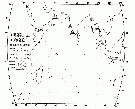 issued from : T.J. Lawson in Marine Biology, 1977, 43. [Fig.5, p.81]. issued from : T.J. Lawson in Marine Biology, 1977, 43. [Fig.5, p.81].
Distribution map for the Indian Ocean. |
 Issued from : M. Bode, A. Schukat, W. Hagen & H. Auel in J. Exp. Mar. Biol. Ecol., 2013, 444. [p.3, Table 1]. Issued from : M. Bode, A. Schukat, W. Hagen & H. Auel in J. Exp. Mar. Biol. Ecol., 2013, 444. [p.3, Table 1].
Dry mass, individual respiration rate from the northern Benguela Current upwelling system along transects at 23°S and 26°40'S, off Walvisbay and Lüderitz (Namibia). |
| | | | Loc: | | | sub-Antarct. (S Indian, S Tasmania, Auckland & Campbell Is., SE Pacif.), New Zealand (Kaikoura, S South Island, sub-Antarct.), Tasman Sea, S Indian (subtropical convergence), South Africa (W), Namibia, SE Atlant., Argentina-S Brazil, Chile, Straits of Magellan, Galapagos-Ecuador (in Tutasi & al., 2011 (p.97, Table 2) | | | | N: | 19 (sub-Antarct.: 2; S Atlant.: 10; Indian: 3; Pacif.: 4) | | | | Lg.: | | | (35) F: 2,55-2,45; M: 2,4-2,34; (36) F: 3,12-2,27; M: 2,84-2,18; (116) F: 2,59; M: 2,45; (202) F: 3; M: 2,03; (432) F: 2,83-2,45; M: 2,55-2,4; (909) F: 2,25-2,7; M: 2,2; 2,35; (1110) F: 2,35-2,74; {F: 2,25-3,12; M: 2,03-2,84}
The mean female size is 2.650 mm (n = 12; SD = 0.2601), and the mean male size is 2.374 mm (n = 10; SD = 0.2219). The size ratio (male : female) is 0.88 (n = 6; SD = 0.1057. | | | | Rem.: | épi-mésopélagique.
Profondeur d'échantillonnage (sub-Antarct.) : 0-100 m.
Voir aussi les remarques en anglais | | | Dernière mise à jour : 28/11/2020 | |
|
|
 Toute utilisation de ce site pour une publication sera mentionnée avec la référence suivante : Toute utilisation de ce site pour une publication sera mentionnée avec la référence suivante :
Razouls C., Desreumaux N., Kouwenberg J. et de Bovée F., 2005-2026. - Biodiversité des Copépodes planctoniques marins (morphologie, répartition géographique et données biologiques). Sorbonne Université, CNRS. Disponible sur http://copepodes.obs-banyuls.fr [Accédé le 27 janvier 2026] © copyright 2005-2026 Sorbonne Université, CNRS
|
|
 |
 |


