Introduction | Morphologie générale | Morphologie des diverses parties du corps et des appendices | Dimensions | Formes | Principales modifications structurelles | Les antennules | Les antennes |
Le cadre buccal : Labrum et labium | Les Mandibules | Les Maxillules ou premières maxilles | Les Maxilles (Maxilla) ou secondes maxilles | Les Maxillipèdes | Les pattes natatoires | Variation du nombre des segments corporels | Nomenclature des parties du corps et Métamérie | Structures et organes tégumentaires | Organes visuels |
Variations de la morphologie | Sex-ratio | Gynandromorphisme et intersexualité | Accouplement | Parthénogénèse | Caractéristique de la ponte |
Les formes larvaires | La durée du développement | La croissance | Régénération |
Anomalies | Classification et Phylogénie | Références | Photothèque
 Morphologie des diverses parties du corps et des appendices : Morphologie des diverses parties du corps et des appendices :
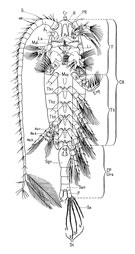
Le céphalosome, en vue dorsale (fig. G), présente en avant un bord libre sur lequel on voit deux petits "cils": les fils rostraux (fr), très discrets, filaments sensoriels ou sensilla. Sur la face ventrale, le front se recourbe en un rostre dirigé vers l'arrière, bifurqué et se terminant par deux pointes, les filaments rostraux (FR).
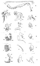
Sur la face ventrale (fig. G2), la tête porte symétriquement, en position préorale, deux appendices plurisegmentés plus longs que le corps, les antennules (A1) (ou premières antennes) (fig. G3 A). Celles-ci sont constituées de vingt-cinq segments plus ou moins égaux chez la femelle (fig. G3, A, a), munies de soies simples ou plumeuses et d'organes sensoriels (les aesthetes ou aesthetasc). Chez le mâle, les deux premiers segments proximaux sont soudés, légèrement dilatés, et portent des organes sensoriels plus développés que chez les femelles. Les antennules sont disposées à 90° de l'axe du corps. G2), la tête porte symétriquement, en position préorale, deux appendices plurisegmentés plus longs que le corps, les antennules (A1) (ou premières antennes) (fig. G3 A). Celles-ci sont constituées de vingt-cinq segments plus ou moins égaux chez la femelle (fig. G3, A, a), munies de soies simples ou plumeuses et d'organes sensoriels (les aesthetes ou aesthetasc). Chez le mâle, les deux premiers segments proximaux sont soudés, légèrement dilatés, et portent des organes sensoriels plus développés que chez les femelles. Les antennules sont disposées à 90° de l'axe du corps.
Postérieurement les antennes (A2) (fig. G3 B, a) montrent une structure d'appendice biramé avec un sympodite ou basipodite (B) composé de deux segments successifs (B1 ou coxa, et B2 ou basis). Sur le basis s'articulent deux branches plurisegmentées: l'une interne, l'endopodite (Enp) à deux segments; l'autre externe, l'exopodite (Exp) formé de sept segments. Tous ces segments sont munis de soies.
La bouche s'ouvre en arrière des antennes, sur la ligne médio-ventrale. Elle est protégée par une lèvre supérieure: le labre ou labrum (Ls), lobe musculaire portant de fines et courtes soies sensorielles. Elle est délimitée à sa partie postérieure par le labium (Li) constitué par une paire de lobes dérivés par fusion des paragnathes.
Les mandibules (Md) (fig. G3 C, a) sont disposées de part et d'autre de la bouche. Elles présentent une structure fondamentalement biramée comportant, une coxa présentant à la base une expansion chitineuse bien développée dont l'extrémité distale montre un processus molaire (pars molaris) qui cache partiellement l'ouverture buccale. Cette bordure masticatrice présente une rangée de dents couronnées de silice. Le basis est réduit, son axe à 90 ° de la lame masticatrice porte un exopodite plurisegmenté à cinq segments et un endopodite à deux segments munis l'un comme l'autre de soies.
Les maxillules (Mx1) (fig. G3 D, a) sont des appendices très modifiés, de forme foliacée à lobes très inégaux externes et internes, armés de soies fortes et fines. La précoxa porte un arthrite (lobe du côté interne) muni de 16 soie fortes; une coxa avec un endite (lobe interne) muni de 3 soies et un épipodite (lobe externe) muni de 9 soies; Le basis porte 2 endites, le proximal (lobe bien individualisé) avec 4 soies, le distal (incorporé au segment) porte 4 soies, et un lobe externe (exite) muni de 1 soie; L'exopodite (en forme de lobe externe à un seul segment visible) porte 11 soies; l'endopodite montre trois segments courts munis chacun de 4, 4 et 7 soies.
Les maxilles (Mx2) (fig. G3 E, a) paraissent uniramés. La précoxa possède 2 endites munis de 6 et 3 soies; la coxa possède 2 endites munis de 3 soies et 1 forte soie externe; Le basipodite est muni d'un d'endite à 3 soies. L'endopodite est à 3 segments (le premier étant fusionné au basis = allobasis) muni de 2 ou 3 soies. Pour Lang (1947) les trois segments distaux sont considérés comme l'endopodite alors que l'unique soie externe serait l'exopodite vestigial. Le nombre des soies des divers lobes est difficile à décompter.
Les maxillipèdes (Mxp) (fig. G3 F, a) sont également des appendices uniramés allongés. Ils appartiennent au premier somite thoracique et sont homologues des pattes natatoires, mais les appendices droit et gauche ne sont pas réunis à leur base. Ils présentent une précoxa; une coxa à 3 endites peu marqués muni de 3, 4, 4 soies; d'un basis muni de 3 soies (du côté médian interne) et du premier segment de l'endopodite plus ou moins incorporé et réduit à 2 soies, suivi de cinq segments munis de 4 soies. Les maxillipèdes sont incurvés ventralement et antérieurement.
Cinq "anneaux" délimités par des sutures dorso-latérales, et la présence d'une paire de pattes biramées, définissent le "thorax" ou métasome. Ils sont comptés d'avant en arrière (Th1 à Th5). Le "dernier" segment thoracique (Th5) présente des angles postéro-latéraux arrondis.
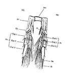 Cinq paires de pattes natatoires sont bien développées ventralement, correspondant chacune à un segment thoracique. Elles sont notées de l'avant vers l'arrière (P1 à P5). Elles présentent toutes la structure type de l'appendice biramé avec une partie basale à trois segments (la précoxa étant peu identifiable) et deux rames à trois segments chacune (fig. G4). Une plaque chitineuse médiane (pm), soudée à la précoxa et à la partie basale de la coxa (à l'exception des Gelyelloida), unit les deux appendices, assurant le synchronisme du battement. Cinq paires de pattes natatoires sont bien développées ventralement, correspondant chacune à un segment thoracique. Elles sont notées de l'avant vers l'arrière (P1 à P5). Elles présentent toutes la structure type de l'appendice biramé avec une partie basale à trois segments (la précoxa étant peu identifiable) et deux rames à trois segments chacune (fig. G4). Une plaque chitineuse médiane (pm), soudée à la précoxa et à la partie basale de la coxa (à l'exception des Gelyelloida), unit les deux appendices, assurant le synchronisme du battement.
Chaque appendice se compose de trois parties (fig. G4):
1- Un basipodite à deux segments: coxa (B1) et basis (B2), la précoxa étant réduite à un sclérite. La coxa présente une soie plumeuse interne, à l'exception de la P5 qui montre un bord interne dentelé. Le basis porte une soie plumeuse interne uniquement dans le cas de P1, et s'articule aux deux rames.
2- L'exopodite (Exp) ou rame externe, comprend trois segments de longueur inégale munis de soies internes longues (Si), d'épines ou soies externes (Se) plus ou moins fortes et courtes selon la patte, et d'une soie terminale (St) plus ou moins développée qui prolonge le segment distal.
3- L'endopodite (End) ou rame interne est à trois segments, plus court que l'exopodite, munis de soies.
Les différences entre les cinq paires de pattes sont peu importantes, mais elles présentent chacune un nombre et une disposition des soies et épines qui leur sont propres donnant lieu à une nomenclature rigoureuse constituant une sorte de fiche signalétique de l'espèce et du sexe, utilisée dans les clés de détermination.
Exemple pour la P4:
Coxa (B1): 0-1; Basis (B2): 0-0; Endopodite (End): 0-1; 0-2; 2-2-3; Exopodite (Exp): I-1; I-1; II- I-5
Les soies (chiffres arabes), et les épines (en chiffres romains), sont comptées de l'extérieur vers l'intérieur pour chaque article, du proximal au distal (Sewell,1949).
La première paire de pattes (P1) est plus courte que la seconde (P2) elle-même légèrement plus courte que les suivantes, la taille maximale est atteinte par la quatrième (P4). La cinquième paire (P5) est quelque peu différente dans les deux sexes et montre une faible asymétrie chez le mâle où l'exopodite de la patte droite est un peu plus courte.
Le corps postérieur (ou urosome), ne porte aucun appendice. Il est de forme cylindrique et s'articule au cinquième segment thoracique (Th5). Il présente quatre segments chez la femelle, le premier (le plus antérieur) est renflé sur la face ventrale, montrant un épaississement recouvrant un orifice génital médian impair. Celui du mâle a cinq segments, avec sur le premier, une petite fente légèrement sur la gauche de la ligne médiane correspondant à l'ouverture génitale.
Le dernier segment de l'urosome, portant l'anus en position dorsale recouvert d'un opercule, est le somite anal, sur lequel s'articulent deux bâtonnets indépendants: la furca ou rames caudales, prolongée distalement par six soies de longueur inégales.
La description et la nomenclature utilisées précédemment s'appliquent dans ses grandes lignes au plus grand nombre des copépodes libres, bien que l'on puisse observer chez ceux-ci de très nombreuses variations dans la forme et la structure du corps et des appendices.
Malgré sa très grande diversité, le groupe dégage une impression à la fois d'unité et de plasticité structurelle qui lui ont permis de coloniser tous les espaces aquatiques marins et dulçaquicoles, mais aussi biologique par leur mode de reproduction (accouplement, et dépôt d'un spermatophore par le mâle sur le "segment" génital de la femelle) et de développement qui comprend outre l'œuf, trois phases présentant une métamorphose (nauplienne, métanaupliénne, copépodite), chacune de plusieurs stades (respectivement et généralement de 3 ou moins, 3 ou moins, 6), le dernier stade copépodite étant l'adulte qui a atteint sa taille maximale, et ne subit plus de mues.
La forme nauplienne, ovoïde ou pyriforme, est caractéristique, présentant généralement un œil nauplien impair médian et trois paires d'appendices: A1 uniramé, A2 et Md biramés.
Les principales modifications structurelles se sont opérées au cours de l'évolution des principaux taxons et à l'intérieur de chacun d'eux sous l'influence de:
1- les habitats qui conditionneront la forme du corps: réductions par fusion de segments du corps antérieur et postérieur, place de l'articulation entre ces deux parties, modification de certains appendices par adaptation.
2- le mode de nutrition en relation avec les sources trophiques disponibles et les modifications plus ou moins prononcées des appendices buccaux.
3- le mode de reproduction qu'impose la séparation des sexes et la nécessité d'un accouplement.
|





 Any use of this site for a publication will be mentioned with the following reference :