



General Morphology of Marine Planktonic Copepods
Visual organs
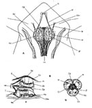
In the great majority of free-living forms there exists an unpaired, median eye corresponding to the eye of the nauplius larva, and therefore commonly called the "nauplius eye". It is generally situated between the antennules on the ventral surface of the head. Its structure was studied by Vaissiere (1961) in Eucalanus elongatus (fig. G15), by Fahrenbach (1964) in a cyclopoid by electron microscopy, and then by Park (1966) who studied the ocular complex in a calanoidan belonging to the family Pontellidae.
It is rendered visible by its coloration due to orange-red, blue or green pigments (Lwoff, 1927). These carotenoid-protein pigments may coexist with black pigment platelets (pro-melanins). The nauplius eye persists unchanged in most Copepods, but in a number of them (Boxshall, 1992) it undergoes considerable transformations that may reach the extreme, as in the bathypelagic calanoid genus Cephalophanes.
The modifications generally consist of a widening of the three ocelli, the doubling of the dorsal ocelli, but above all the appearance of translucent cuticular dioptric lenses, as in the family Pontellidae, the Miracidae (Harpacticoida), the Sapphirinidae and Corycaeidae (Poecilostomatoida).
‹ Tegumental structures and organsMorphological variations ›