



General Features of the External Morphology of Marine Planktonic Copepods
The buccal frame: Labrum and Labium
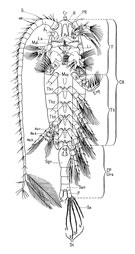
The frame delimiting the oral atrium presents a complex structure related to the feeding mode, which can strongly modify the appendages contributing to it.
A detailed description of the buccal frame was provided by With (1915).
Given the types of habitat and the diversity in time and space of available trophic resources, adaptations inevitably lead to varied structures. Thorell (1859) had already relied on these structures to define three groups among Copepods, which could form the basis of a natural classification.
1 The Gnathostomes, which have an oral cavity delimited by a large labrum and a small labium (paragnaths) and covered laterally by the masticatory plates of the mandibles and their gnathobases. They are adapted for chewing.
2 The Poecilostomes, which have oral appendages similar to the former but greatly reduced, and adapted for brushing or licking fine particles.
3 The Siphonostomes, which have a tubular oral cavity formed by the more or less complete fusion of the labrum and labium, and whose mandibular gnathobases constitute stylets. They are adapted for sucking.
One can thus see how the group has diversified, transitioning from free-living forms to commensal or semi-parasitic, and then fully parasitic forms.
While these structures are of great taxonomic importance, they may also be misleading due to possible adaptive convergences. For Sars (1903), the existence of gradual transitions between these three groups, and the arbitrary separation of forms sharing other common characters, do not allow buccal structures to play a primary role in high-level taxonomic groupings.
The labrum and labium form the anterior and posterolateral borders of the mouth and are not considered as appendages.
The labrum, situated at the level of the A2, constitutes a median muscular lobe on the anterior border of the mouth, furnished with external spinules and pores of the labral glands on its posterior surface (Arnaud et al., 1988).
The labium consists of a pair of separate lobes, derived from the paragnaths (sternal structure), ornamented with a row of spinules and sometimes fine setae.
The shape and dimensions of the labrum and labium become more complex, slightly so in the Cyclopoida and Poecilostomatoida, but considerably more in the Siphonostomatoida (Huys & Boxshall, 1991, p.343), forming a laterally split cone into which the mandibles are inserted; the paragnaths are fused to form the labium, which acts as the lower lip of the oral cone.
Thus the structure of the mouth justifies the distinction between the Poecilostomes and the Siphonostomes, and the disappearance of the former order Cyclopoida as established by G.O. Sars.
In the Calanoida and in most other orders, the labrum (anterior to the buccal frame) appears as a muscular median lobe, capable of slight movement in the anteroposterior plane (Boxshall, 1985). It often presents rows of spinules anteriorly and posteriorly, and bears pores of labral glands on its posterior surface. The paragnaths are paired lateral lobes (or labium) bearing rows of spinules.
In the Poecilostomatoida, the mouth is a transverse slit; the anterior border of the slit is overarched by the labrum (a flattened structure of variable shape), while the posterior border is rectangular, often concave, sometimes bearing denticles. In some commensal species the labrum shows sexual dimorphism (Ho, 1970). The lateral sides of the mouth are open to admit the oral appendages. The lower border of the mouth is delimited by the labium, a structure that is often poorly defined, appearing formless, sometimes as a protruding lobe. This labial swelling is lower than the labrum. It has been suggested that, in this group, the primitive feeding mode involves passage of nutritive particles from the front towards the oral orifice. This would functionally explain the low position of the labium above the ventral surface of the cephalothorax.
The mouth of the Cyclopoida (stricto sensu) is essentially similar to that of the Poecilostomatoida.
In the Siphonostomatoida, the labrum and labium, partially or completely fused, form a tubular cone or siphon above the opening of the oesophagus. The lateral borders of the lips, when they remain separated from each other over a short distance at their base, allow the mandibles to penetrate inside the oral cone (Kabata, 1979).
In the Monstrilloida, a discrete oral cone is visible on the more or less anterior ventral surface of the cephalosome. It terminates in a pore (Huys & Boxshall, 1991).