


General Features of the External Morphology of Marine Planktonic Copepods
The swimming legs
Their number and shape will depend on the fusion mode of certain somites, as well as on sexual characteristics.
The number is a function of the tagmosis of the genital region, which affects the posterior thoracic segments.
The thoracic appendages are fundamentally swimming appendages, but, as previously noted, the cephalisation of the first thoracic somite has brought about a modification of the first pair into maxillipeds. The second pair may also be affected, albeit to a lesser degree. The same applies to the two pairs of posterior thoracic appendages, which are highly variable among groups and between sexes.
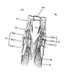 The structure, according to the scheme of Hansen (in Gurney, 1931), is reducible to the primitive biramous appendage. It comprises three basal segments, the first or Precoxae being poorly identifiable, reduced to a sclerite at the base and laterally to the second segment. The latter, or Coxa (formerly designated as basipodite 1), bears, like the following segments, an internal seta and an external spine or seta, which, given the fusions or modifications of segments, appears as an archaic character that can be used as an index of the evolution of forms. The Basis (formerly basipodite 2) bears an inner ramus (Endopodite) and an outer ramus (Exopodite), each with at most three segments (fig. G4).
Gurney (1931) attempted to establish homologies between the segments of copepod swimming legs and those of primitive malacostracans. In total, nine segments can be identified if one accepts the fusion of the precoxae and pre-ischium, and the fact that in the distal segment of the endopodite and exopodite, the internal setae in the former and the external spines in the latter delimit segments, thus defining four fused segments.
The legs of each pair, at least the first four (maxillipeds excluded), are connected by the inner lateral borders of their coxae via a strong sclerite forming an intercoxal plate, which imposes synchronous beating of the right and left legs. This basal plate constitutes an apparently unique structure (Manton, 1977) that gave this group of Crustacea its name Copepoda (from the Greek Kopion: small oar, and podos: foot).
By virtue of their function, the various pairs of swimming legs show great similarities in a single individual, but none, owing to the fusion of certain segments and their setal armature, is exactly identical to the preceding or following pair. This characteristic is used in faunal studies as one of the criteria for species determination.
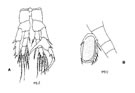
The great majority of free-living forms have only five pairs of legs; the fifth, adjoining the genital tagma, is in many cases non-natatory, profoundly modified, either by reduction, which can even lead to their complete disappearance, or conversely hypertrophied and asymmetrical, in males only (fig. G12, A).
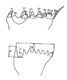 This change in function, with the corollary of a structural modification, is manifested in the male (very exceptionally in the female of a harpacticoid: Phyllopodopsyllus) (fig. G11, B), introducing a particular dimorphism of the appendage (frequently used in faunal studies). This occurs when the fifth pair of legs (maxilliped excluded from the numbering) of the male is adapted for involvement in reproductive activities: grasping the female, locating her genital area, and manipulating the spermatophore.
In certain copepods, and very generally in the groups Harpacticoida and Cyclopoida, the fifth pair is greatly reduced and its parts are difficult to interpret. It consists of a short basal part, laminar or subcylindrical, bearing one or more setae or spines. On either side of the oviduct openings, a sixth pair of legs is sometimes observed, even more reduced than the previous one, forming two projections on either side of the median axis, bearing setae.
The loss of function of this sixth pair of legs would have permitted the use of the muscles for the genital operculum, itself a remnant of a pair of legs that closes the genital openings (Huys and Boxshall, 1991). Swimming legs of normal construction were undoubtedly incompatible with the attachment of the egg sac(s) to the genital tagma, and with the presence of the spermatophore.
Anteriorly, the first pair of swimming legs (P1 in conventional nomenclature) shows variations that may involve a slight reduction in size relative to the following pairs and a reduction of the endopodite. Furthermore, this first pair of legs may undergo structural changes that make it resemble the maxillipeds.
In Harpacticoida, Lang (1948, p.64) provides a table of comparative figures that are illustrative of these variations.
Similar changes occur, although more rarely, in the second pair of legs (P2), for example in the harpacticoid Canthocamptus. According to Kabata (1979), these modifications occur in forms that already possess prehensile appendages (A2, Mxp) and serve to increase attachment capacities.
The study of locomotory behaviour in relation to the environment, which is often poorly understood, is nonetheless indispensable for understanding the variation in structures.